Dye doped spectroscopy interpretation

Join the discussion
Ask a follow-up here, or get your own question answered by working scientists, mathematicians and engineers — people, not an autocomplete.
Real named experts · corrections over time · the nuance an AI answer skips
8 replies · 2K views
Srv44
Messages
3
Reaction score
0
Hello all,

I am working on a dye doping project and got a photospectrometry result as show in a picture for my thin film. Can anyone please help me interpret the physical meaning of the graph below? Thanks!
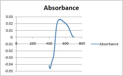
upload_2016-7-20_11-58-1.png
 
Engineering news on Phys.org
Negative Absorbance, the same as measuring light that is either generated by something other than the Spectrometer (reflections from an outside source?); or equivalently the calibration is incorrect.
 
Your sample is very dilute with an absorbance of 0.03 AUFS. Most dyes have incredibly high molar absorption values in the visible range. Did you zero your spectrophotometer over the entire wavelength range? What is your solvent?
 
Kevin McHugh said:
Your sample is very dilute with an absorbance of 0.03 AUFS. Most dyes have incredibly high molar absorption values in the visible range. Did you zero your spectrophotometer over the entire wavelength range? What is your solvent?

Kevin, yes I did zero the spectrophotometer, and the sol I used is made up of : Tetraethyl orthosilicate, Ethanol, hydrogen phosphate and water.
 
You mention a thin film, is your sample a solid or a liquid? Are you using quartz cuvettes, or a holder for solid thin film?
 
Kevin McHugh said:
You mention a thin film, is your sample a solid or a liquid? Are you using quartz cuvettes, or a holder for solid thin film?

My sample is a solid, so a film of dye coated on a microscope slide. Thats all it is. While getting the spec data, I place the dye coated slide right on the beam path against the cuvette holder. Is that something that affects the data I get?
 
Either the glass slide or your sample may fluoresce. I've had cheap photograph lens filters fluoresce Red when hit with intense Blue light. Most annoying.
 
You probably need to use quartz. Are you using a dual beam instrument? Also, your path length will be variable, no good for quant work. How are you zeroing the spec? Your baseline looks like the machine isn't comparing similar backgrounds. Using quarts cuvettes, with dye in solution at known concentration is your best bet to successful spectroscopy. Increase your concentration to achieve at least 1 AUFS for best results. Use methanol as your solvent, it is clear to 205 nm.

Edit: Belay the last, I see your casting a thin film. I believe there sample holders that use mm scale spacers for thin film work in solution. You would do well to get them. Use quartz.
 
Last edited:
  • How do the orthosilicates and the phosphate affect the UV-Vis spectrum? I would expect them to be optically clear, but there may be some other phenomenon going on between the adjutants and the dye.And, as Tom said, does your sample fluoresce? What does the spectrum of the pure dye in ethanol/water look like compared to in solution with the orthosilicates and phopshate>