Is concentric helix DNA sculpture valid?

  • Thread starter Thread starter DaveC426913
  • Start date Start date
  • Tags Tags
    Dna Shapes
Join the discussion
Registration is free. Ask a follow-up in this thread, or start your own.
12 replies · 3K views · last reply
DaveC426913
Gold Member
2025 Award
Messages
24,754
Reaction score
9,175
TL;DR
Are both these valid representations of the DNA double helix?
I'm designing a ... let's call it a sculpture. It represents DNA.

I just realized there are (at least) two ways helices can spiral together:

Two side-by-side diagrams of ribbon helices with red dotted axis lines: A shows two helices with different radii, in phase; B shows two identical helices offset by 180 degrees.


A has two helices that are concentric but with different radii. They are also "in phase".
B has two identical helices that are "out-of-phase" by 180 degrees.

A is what I started with - and what I want - but I don't know if it is a valid representation of DNA (if you uncurled it, the inner helix is shorter than the outer helix. It would curve around to form a "wagon wheel" pattern.

Or am I being persnickety? After all, if DNA is bendy enough, and if the radii in A were long enough, in theory, the length disparity would effectively disappear. In other words, sufficient unfurlification of B would result in A.

So, my question is: is A a reasonable representation of DNA - i.e. perhaps odd but not wrong?
 
Attachments
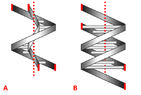
  • Two side-by-side diagrams of ribbon helices with red markers and a dotted vertical axis; A shows two helices of different radii in phase, B shows two identical helices 180 degrees out of phase.
    1666584051033.png
    25.1 KB · Views: 166
Chemistry news on Phys.org
Its more like B than A.
However, there is a major groove and a minor grove, like this:

Space-filling model of B-form DNA showing the major and minor grooves, labelled, with red and green zigzag lines tracing each groove along the double helix.

The backbone parts are charged and coil around the outside.

Diagram of DNA major and minor grooves: a space-filling model of a double helix on the left, with base pairs labelled A, D and A, linked by black lines to molecular structures of adenine, thymine, guanine and cytosine on the right, with coloured arrows marking hydrogen-bond sites.


The backbones of the two strands go in opposite directions chemically along the molecule (anti-parallel). Probably doesn't matter to your model though.
 
  • Informative
  • Like
Likes   Reactions: berkeman and Lord Jestocost
DaveC426913 said:
TL;DR Summary: Are both these valid representations of the DNA double helix?

I'm designing a ... let's call it a sculpture. It represents DNA.

I just realized there are (at least) two ways helices can spiral together:

View attachment 315948

A has two helices that are concentric but with different radii. They are also "in phase".
B has two identical helices that are "out-of-phase" by 180 degrees.

A is what I started with - and what I want - but I don't know if it is a valid representation of DNA (if you uncurled it, the inner helix is shorter than the outer helix. It would curve around to form a "wagon wheel" pattern.

Or am I being persnickety? After all, if DNA is bendy enough, and if the radii in A were long enough, in theory, the length disparity would effectively disappear. In other words, sufficient unfurlification of B would result in A.

So, my question is: is A a reasonable representation of DNA - i.e. perhaps odd but not wrong?
Form A does not appear in real life.
 
  • Like
Likes   Reactions: DaveC426913 and BillTre
Hornbein said:
Form A does not appear in real life.
I am amazed that such a thing is theoretically possible (though pardon me that I'm not 100% confident in your assertion).
 
Hornbein said:
I am amazed that such a thing is theoretically possible...
Not sure what you mean. 'splain?
 
DaveC426913 said:
Not sure what you mean. 'splain?
? I dunno, it's just a very weird possibility.
 
Hornbein said:
? I dunno, it's just a very weird possibility.
Oh, you mean impossible specifically for DNA.
 
One of the major problems with the "A" version would be the location of the sugar-phosphate backbone. It has electrical (static) charges. The bases (nucleosides, nucleotides include bases sugars and phosphates) are not or only weakly charged. The two strands will be most stable with the charges as far a apart as possible within the molecule. Thus, the two backbones would be as far apart as possible. the bases that do the pair bonding are in the middle snuggled up to each other.

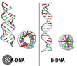
The B form of DNA is what people usually think of, but there are other forms:
A-DNA: twists in the same direction as B-DNA, but the base orientation is idfferent
there are also other weird forms or DNA possible. there is an
Z-DNA: twists in the opposite direct from A and B-DNA. I think this form depends on certain sequences. It is definitely affected by supercoiling of the double helix (which puts twisting tension on the molecule).
Three DNA structure diagrams labelled A-DNA, B-DNA and Z-DNA, each showing a side view of the helix with a view down its axis beside it; base pairs are coloured red, green, purple and cyan.


Just to make things more confusing, there are also some 3 and 4 strand DNA structures:

Three ribbon models of helical nucleic acid structures labelled Duplexe, Triplexe and Quadruplexe, shown left to right with two, three and four intertwined strands respectively.
 
Cool.
That answers a question I had when I saw this:
Space-filling model of B-DNA double helix with blue and tan atoms, red and green zigzag lines marking alternating major and minor grooves, labelled Minor groove and Major groove.

I wondered how this was possible, if both strands were identical and the structure were axially symmetric. I started thinking about what it would do to the nucleotides.

But this shows how it's possible:

Ribbon-and-stick model of a double helix DNA structure, with a grey sugar-phosphate backbone and horizontal base pairs in red, green, blue and magenta.


The two ends of the nucleotides are staggered along the length (the vertical axis). That's what leads to the alternating major/minor groove. Right?

TIL.
 
Attachments
  • Side-by-side ball-and-stick and ribbon models of A-DNA on the left and B-DNA on the right, each with a view down the helix axis, labeled A-DNA and B-DNA.
    1666666783695.png
    44.8 KB · Views: 204
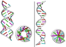
  • Two 3D models of DNA double helices shown side by side, with grey backbones and short colored rods for base pairs, plus end-on views of each helix showing the base pairs arranged in rings.
    1666666765219.png
    41.6 KB · Views: 208
DaveC426913 said:
Oh, you mean impossible specifically for DNA.
Yeah, incompatible with its function.
 
OK, more stuff.

This diagram
Space-filling model of B-DNA showing a double helix with blue and gold atom spheres; red and green lines mark the minor groove and major groove on each side.

indicates that the nucleotide pairs span the minor groove, not the major groove.

I'm interested to know what the "phase" difference of the two strands are. If it were 180 degrees, the two gaps would be identical.

More to-the-point, I'm interested in knowing what the arc (of a full circle) of a given nucleotide pair is.

This diagram suggests that it might be around 135 degrees.

Two views of B-DNA: at left a double helix with coloured base pairs, at right the same structure viewed end-on showing base pairs radiating from the central axis with a label reading B-DNA.

Side view of a B-DNA double helix on the left, and on the right an end-on view of the helix with colored base pair rods radiating from the center, with a red line drawn across it; caption reads B-DNA.

I'm not looking for exact numbers, just the gist.
 
DaveC426913 said:
OK, more stuff.

This diagram
View attachment 316653
indicates that the nucleotide pairs span the minor groove, not the major groove.
Aha that explains it. It's hard to see that.

DaveC426913 said:
I'm interested to know what the "phase" difference of the two strands are. If it were 180 degrees, the two gaps would be identical.

More to-the-point, I'm interested in knowing what the arc (of a full circle) of a given nucleotide pair is.

This diagram suggests that it might be around 135 degrees.

View attachment 316654
View attachment 316655
I'm not looking for exact numbers, just the gist.

I'd call that the pitch of the helix. Not that I'm any expert.
 
Nope. Turns out I was completely wrong and the diagram is highly misleading.

From the specific angle of the diagram, the two helices are exactly 180 degrees out of phase. It only looks like the helices are unevenly-spaced:

Space-filling model of B-DNA showing two sugar-phosphate backbone helices with labelled major and minor grooves, with magenta measurement lines marked 180px and red and green guide lines tracing the strands.


And the asymmetry is not about the nucleotides either. Here is an outline of the helices - the nucleotides do not produce the asymmetry in the grooves - at least, not at this angle:
Space-filling model of B-form DNA showing two backbone helices with the major and minor grooves labelled, with blue phosphate and sugar atoms wrapped around beige base pairs.


But if I measure the helices at a point 90 degrees rotated, you can see that they are not even; it's a 5:7 ratio.
Space-filling model of B-DNA showing two intertwined helices with labelled major and minor grooves, with pixel measurements 210px, 150px and 210px marked along a horizontal blue line and diagonal guide lines.


This suggests that the major/minor groove is a product of the bumpiness of the helices themselves.