Prof. Verschure's thin section collection from the Fen and Hor localities

185 replies · 71K views · last reply
Science Advisor
Education Advisor
Insights Author
Messages
7,852
Reaction score
4,227
TL;DR
petrology
(Edit: since the thread title was changed, this first sentence is too cryptic: the original title referred to a Tool song....)

Besides being a favorite song by a favorite band, the thread title is a straightforward play on words. This summer, as a present to myself for being promoted, I purchased a collection of thin sections that I believe comprise the research materials of Prof. Rob Verschure, who at the time was faculty in the Geological Institute in Amsterdam.

What changed this purchase from eccentric (although, at $2 per sample, also very affordable) to something more elevated is that Prof. Verschure published his findings on many of these samples, primarily in:

https://www.ngu.no/FileArchive/NGUPublikasjoner/NGUnr_380_Bulletin_70_Verschure_35_49.pdf

and

https://research.vu.nl/en/publicati...-parameter-for-metasomatism-and-its-applicati

i.e. I have a "Rosetta stone" for explosion breccias, carbonatites, and damtjernites. Many of the collected samples have been fully characterized, for example this thin section:
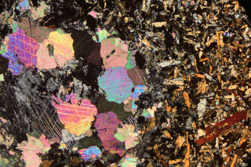
Thin section photomicrograph under cross-polarized light showing a coarse-grained rock with colourful interference-coloured crystals in pink, magenta, blue and yellow, set against a fine-grained dark matrix of elongated orange and grey crystals.


Sample Hor 1 has been classified as a calcite-bearing clinopyroxene-hornblende lamprophyre that has been dated to 313 Ma. This sample contains abundant augite and brown hornblende.

This sample (Fen 23) consists of zoned augite (I think...) and carbonates, dated to 594 Ma:
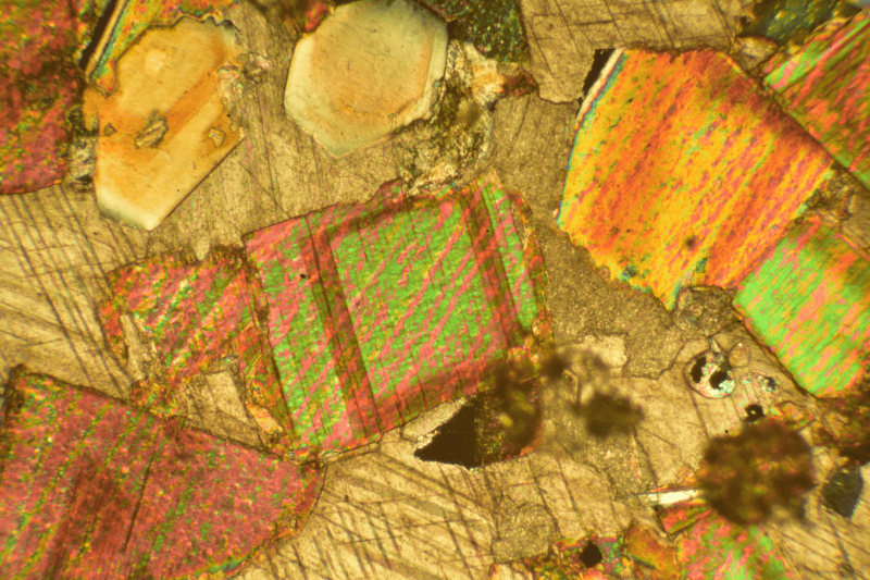
Thin section photomicrograph under cross-polarised light showing interference-coloured mineral grains in pink, green, yellow and orange, with a grey-brown matrix and dark opaque inclusions.


Many of the samples are carbonatites, but there are also many examples of schists and gneisses; here's a schist with pronounced folding:
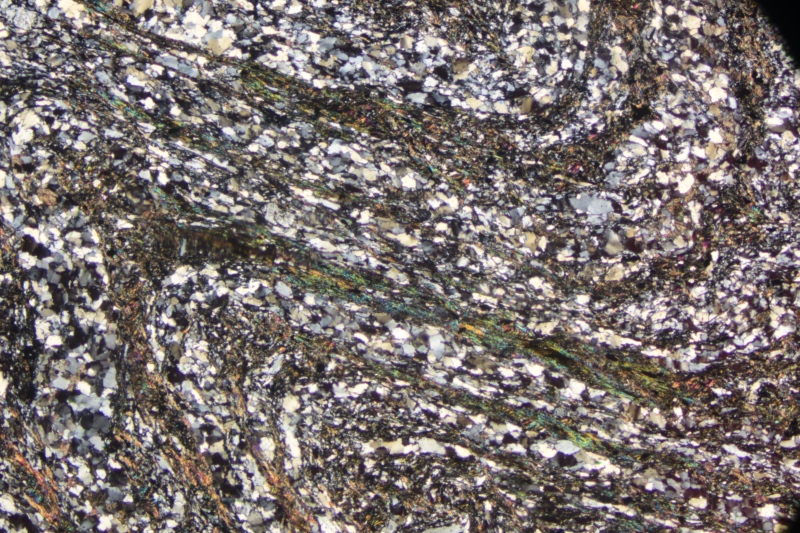
Cross-polarised light photomicrograph of a metamorphic rock thin section showing elongated mica flakes with interference colours wrapped around granular quartz and feldspar grains.


I'm hoping to learn quite a bit as I get more familiar with the variety of samples...
 
Attachments
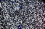
  • Thin section photomicrograph under crossed polarized light, showing a dense mosaic of small grains in bright white, grey and dark tones with scattered deep blue patches.
    1690565551812.png
    313.9 KB · Views: 243
Last edited:
  • Like
Likes   Reactions: WWGD, Spinnor, sbrothy and 10 others
I realized that while the microstructure of these rocks are interesting, the mesoscopic structure is often more photogenic- here are some samples photographed at 1:1 using a 55mm Micro Nikkor:

Thin section of rock on a glass slide viewed under cross-polarised light, showing an interlocking mosaic of colourful mineral grains, with a fine-grained pale band through the centre and a handwritten label reading 34 HAO 2.

Photo of a rock thin section on a dark slide, backlit under crossed polarisers, showing a grey-brown granular matrix with a bright blue and white crystal cluster at centre and a handwritten label on the right reading 94 Hf 1.

Photomicrograph of a thin section under crossed polarised light showing a mosaic of brightly coloured interference-colour crystals in a grey matrix, with a handwritten-style label reading 167 FEN 23 on the mount.

Thin section of a rock mounted on a glass slide under cross-polarised light, showing a fine grey matrix with scattered coloured grains and black opaque fragments; handwritten label reads 9S-FEN 260.


The other advantage is that I have the label in the image so I can more easily keep track of what I want to image at higher magnifications....
 
  • Like
  • Wow
Likes   Reactions: Spinnor, sbrothy, pinball1970 and 4 others
Here's my first 'official' presentation of one of the samples:

Thin section photomicrograph under crossed polars of a fine-grained groundmass with dark rounded clasts and scattered blue crystals, cut by pale carbonate veinlets running diagonally.


This is an example of a "Tveitan carbonatized damtjernite-like explosion breccia". It was obtained from UTM coordinates 5356-65419, is 3.71% Potassium, 0.0888 ppm Wt of radiogenic 40Ar (5% of which is 'atmospheric' 40Ar) and was dated to 316 Ma. From the paper: "[The breccia consists of] a wide variety of xenoliths and xenocrysts in a very fine-grained groundmass consisting mainly of carbonate, green biotite, opaques, and apatite. [...] Many of the xenoliths and xenocrysts are strongly altered, but the abundant apatite phenocrysts and the cores of biotite phenocrysts and perthite xenocrysts do not show any alteration. Numerous veinlets of carbonate transect both the xenoliths and the groundmass"

The primary feature (I would call it a 'phenotype') I have come to associate with damtjernite are the rounded masses ("pelletal lapilli"). Here are a couple of higher-magnification views of this feature (crossed polars):

Thin section photomicrograph under crossed polars showing a rounded, fine-grained pelletal lapilli with a greenish-brown groundmass and small bright grains, cut by a pale carbonate veinlet running diagonally across the field.


and, from a different sample:

Thin section photomicrograph under crossed polars showing a rounded pelletal lapillus with a fine-grained, brownish-orange interior and pale rim, set in a dark green groundmass with coloured mineral grains.


The origin of these has (apparently) been a longstanding mystery- I found this paper claiming to 'understand' them as 'fluidised spray granulation':

https://www.nature.com/articles/ncomms1842

definitely not intro geology!
 
  • Like
  • Informative
Likes   Reactions: sbrothy, pinball1970, DennisN and 2 others
Next up, I was able to reproduce a few images appearing in:

Verschure, R. H. & Maijer, C. 2005: A new Rb-Sr isotopic parameter for metasomatism, ∆t, and its application in a study of pluri-fenitized gneisses around the Fen Ring Complex, South Norway. Norges geologiske undersøkelse Bulletin 445, 45–71.

Here's a piece of the published image (this is an open-source paper, btw.)

Four labeled photomicrographs (c, d, e, f) of thin sections in plane-polarized light; c shows a yellowish biotite grain with dark reaction rim, d shows a pale grain with radiating dark needles, e shows green and yellow grains in a rim-like pattern, f shows a dark grain with striations. Scale bars read 200 µm, 200 µm, 200 µm and 100 µm.

and mine (I have 3 of the 4 above samples):

Photomicrograph in plane-polarised light of a yellowish-brown biotite grain with a dark green reaction rim at its edges, surrounded by pale quartz.


Photomicrograph in plane-polarised light of a biotite flake with pale yellow-brown pleochroism, its edges rimmed by dark radiating needle-like crystals and a small opaque black grain at the lower left.


Photomicrograph in plane-polarised light of a yellow-brown biotite grain with fine dark cleavage lines, adjacent to clear quartz and dark, altered reaction-rim material.


Here's the relevant part of the figure caption:

"Fig. 10. Photomicrographs of fenitized gneisses. […].

(c) Ma 88: Biotite with reaction rim of massive Na-pyroxenes and alkali feldspar due to Fenitization-1. The rim was formed where biotite was in contact with quartz (clear) and not where biotite was in contact with feldspar (turbid). Other biotites within the same sample (not visible on this photomicrograph) appear stronger or even completely replaced. Location of the sample: Small quarry for road material near Steinsrud on road Holla-Steinsrud, Økonomisk Kart Foreløpig Utgave 1971 (BV 030-5-4) coordinates: 51502- 656890.1.

(d) Fen 125: Biotite replaced during Fenitization-2 by radiating needles of bluish Na-amphibole, subsequently partially replaced by fine- grained magnetite and hematite. The replacement occurred where biotite was in contact with quartz. Other biotites within the same sample appear unaffected or completely replaced. The sample was taken 1750 m south of the Fen Complex, far outside the zone of Fenitization–1. Location of the sample: HSP post M00059, Økonomisk Kart Foreløpig Utgave 1971 (BW 020-5-1) coordinates: 138965-51753. […]

(f ) Fen 33’’’: Biotite with a rim of a massive aggregate of blue Na-amphibole pseudomorphing Na-pyroxenes. Stilpnomelane (brown radiating flakes) occurs at the edge of the blue Na-amphibole aggregates. Location of the sample: Håtveittjørn Section 375 m from the contact with the Fen Complex. "

For me, this kind of information is really useful- not for the technical detail- I don't yet have enough knowledge to fully appreciate them- but rather (1) I learn how a geologist looks at samples, and (2) for the experience observing how color is used as a identification tool- I can't rely on color names to identify the mineral. For example, now when I read "bluish Na- amphibole", I understand what that color looks like to my color-deficient eyes. That holds especially for green/brown minerals.

Also, "stilpnomelane" :) .

Photomicrograph in plane-polarised light of a dark green, fibrous biotite grain with a ragged margin fringed by yellow-green and orange reaction rim material against a pale background.
 
This sample, "TVE 15", was obtained from UTM coordinates 5356-65419 and is classified as a "sheared gneiss":

Thin section photomicrograph under crossed polarizers of a sheared gneiss, showing a granular mosaic of blue and white grains with elongated, streaked quartz. Handwritten label reads TVE 15.


(All images are taken with crossed polarizers). This sample is one of a group of "crustal xenoliths from the carbonatized damtjernite-like explosion breccia near Tveitan, Bamble region". This particular sample is considered "country rock", rock that was already present during the explosion event and is dated to 1.4 Ga based on 87Sr/86Sr measurements.

It's clear there is something unusual about this sample- notice the elongated bright 'streaks' throughout the rock. These are sheared/strained quartz grains:

Thin section photomicrograph under crossed polarizers of a sheared gneiss, showing elongated grey and bright quartz and feldspar grains aligned in a band, with dark matrix between them.


Note the presence of 'slip planes', which are visualized as undulose extinction. These generally lie parallel to the strain direction. The major components of this sample are quartz, K-feldspar (perthite) and plagioclase. Much of the plagioclase feldspar has undergone either sericitization or albitization. Minor amounts of biotite are also present (for example, there's a small biotite crystal located below the center of the second image).

I tried to calculate the pressure required to deform the quartz crystals, assuming the crystal started as a cuboid and was deformed via shear, but I got numbers that are way to large- about 106atm. Clearly, slip planes reduce the yield stress and there are other considerations I am yet unaware of.

In any case, I started thinking about grain deformation/recrystallization dynamics and wondered about feldspar grains- quartz always has clearly demarcated grain boundaries, but feldspar does not seem to. For example, look at the (microcline) feldspar grains located just below the smaller deformed quartz crystal, left of center:

Cross-polarized thin section micrograph of a sheared gneiss showing elongated quartz grains with undulose extinction, interlocking with plagioclase and K-feldspar grains in shades of blue and grey.


There are two bright-ish 'arcs' that separate the grains, and I have seen this particular pattern repeated in other samples. A closer look shows that the boundaries between feldspar crystallites have a much different appearance than quartz:

Blue-tinted thin section photomicrograph under crossed polarizers showing quartz grains with a bright streak, undulose extinction and a dark boundary between grains.


In contrast, neighboring quartz grains are (almost) always separated by a dark line:

Blue-tinted photomicrograph under crossed polarizers of a thin section showing interlocking grains, with a small bright grain cluster near the centre and a dark opaque streak at the upper right.
The last image is of multiple microcline crystallites with a large feldspar grain that has undergone sericitic alteration, also showing symplectic alteration (symplectite):

Cross-polarized thin section photomicrograph of a sheared gneiss showing quartz and feldspar grains with blue interference colours, bright elongated streaks and wavy undulose extinction bands.


Does anyone know of a good reference that discusses various grain deformation/recrystallization processes? The next sample I want to show has me thoroughly confused....
 
  • Like
  • Love
Likes   Reactions: sbrothy, pinball1970, DennisN and 1 other person
Today's sample is "Fen 59", mostly because I get to use a bunch of weird words....
Thin section photomicrograph of a carbonatite sample under cross-polarised light, showing pale grey carbonate matrix with dark and blue-tinted crystals; label reads 69 Fen 59, 600 mm.


This carbonatite sample was obtained from the Håtveittjørn section of the Fen complex, an inferred distance of -230m from contact with the country rocks... not sure how to interpret a negative distance. As I mentioned, this sample was a little confusing. At 1:1 zoom of the above image, it's clear that most of the material is a carbonate (for example, calcite and dolomite), but there are also what appear to be numerous subhedral and anhedral oikocrysts enclosing (most likely) anhedral carbonate chadacrysts:
Cross-polarised thin section photomicrograph of a carbonatite: a fine mosaic of grey and bright interference-coloured carbonate grains with scattered dark blue-black rounded and irregular grains.


What little I have read about pokilitic textures typically state that chadacrysts are euhedral, so one confusion that I still have is understanding how these formed. The other challenge was identifying the oikocryst mineral: neither quartz nor feldspar nor pyroxene nor garnet nor leucite. here are PPL (parallel polarizers) and XPL (perpendicular polarizers) views using a 4x objective:
Cross-polarised thin section photomicrograph of a carbonatite showing a poikilitic texture: rounded clear oikocrysts, likely apatite, enclose many smaller grey carbonate chadacrysts, with brownish carbonate matrix.


Cross-polarized thin section micrograph of a carbonatite: rounded dark-blue to black oikocrysts with carbonate chadacrysts set in a pale grey-cream carbonate matrix.

In the PPL image, I decentered the illumination to improve the relief. The carbonate is grey and the oikocrysts are clear and in higher relief. It took some effort, but I finally identified the oikocrysts as apatite- the appearance of apatite in thin section for every kind of rock *other* than a carbonatite is "small crystals, hard to detect". Note also the carbonate vein and the small green/brown (remember- I don't do colors very well) crystals.

A closer look at some of those smaller crystals (surrounded by carbonate) in PPL and XPL is next. Make note of the birefringence of the crystals, the crystal on the left edge that is clear, and the corona of very small crystallites throughout:

Thin section photomicrograph in plane-polarised light showing a poikilitic texture: rounded, anhedral carbonate grains (grey) enclosed by larger clear oikocrysts with higher relief.


Cross-polarized thin section micrograph of carbonatite showing large birefringent crystals with bright interference colors enclosing grey carbonate grains in a poikilitic texture.


So now we go to the relevant paper. The composition of this rock was reported as 60% carbonate, 15% biotite, 20% apatite (confirmation!), 1% quartz, 4% chlorite, and accessories of opaques, rutile, allanite, stilpnomelane, and dispersed carbonate. Since the crystals above are clearly not biotite, there's more to the story: "Fenitization"- an alkali metasomatic event.

In the "Remarks" data column, this rock is characterized as "MF2carbonatite; Bt ⇒F2Chl, F2Qtz,F2Carb rims", and now I can understand what I am looking at: a carbonatite, moderately Fenitized twice.

The original biotite, which appeared 583 Ma ago during the volcanic event, was subsequently altered first by a metasomatic Fenitization (F1) event that broke down the biotite, and then altered again (Fenitization-2), a late hydrothermal process replacing the F1 minerals, producing new fine-grained crystals of chlorite, quartz, and carbonates. And indeed, in the images it can be seen that the biotite crystals were replaced with chlorite (preserving the micaceous habit) and quartz, and also contain a reaction rim consisting of small carbonate crystallites.

Phew! That's a lot. Just getting warmed up, tho....
 
  • Haha
  • Like
  • Wow
Likes   Reactions: sbrothy, Tom.G and DennisN
This, according to the paper, is a sample of 'melteigite', obtained from the Mjølteig A section of the Fen complex, -114 meters from country rock:

Thin section of a fine-grained, multicoloured rock under cross-polarised light, showing many small interference-coloured mineral grains on a dark background; label reads '94 FEN 243'.


One thing I am learning is that the names of these 'exotic' rocks are often in flux- another name for melteigite is 'nephelinolite'. Melteigite comprises one end of the ijolite series, a type of plutonic rock consisting primarily of cliopyroxene and nepheline. Melteigite is at or near the melanocratic end, with the lowest fraction of nepheline while urtite, the most leucocratic, has the highest fraction of nepheline.

This sample is 'officially' described as a "strongly fenitized (F2) meltegeite, with coarse-grained zonal aergirine with titanaugite coronas". Without this, I would have absolutely no way to make sense of the images. For example, here's a pair (PP and XP) using a 4X objective:

Photomicrograph in plane-polarised light of a thin section showing mostly green pleochroic pyroxene crystals with dark, irregular boundaries and colourless striated grains at right and upper left.


Photomicrograph of a thin rock section under crossed polarized light, with vivid interference colours in blue, green, yellow and magenta across densely intergrown mineral grains.


I can maybe recognize a zoned-aergirine crystal in the center and (probably) a Ti-Aug crystal in the lower right. Other than that....???

Here's an image pair taken with a 16X objective from a different location:
Thin section photomicrograph in plane-polarised light showing a rounded grey-brown crystal with dark irregular margins and fine dark cracks, set in a pale green-grey matrix with scattered dark opaque specks.

Photomicrograph of a thin section in cross-polarized light showing a rock with interference colours: a blue-purple zoned crystal and yellow-green grains surrounded by bright blue and white mineral aggregates.


I'm slightly more comfortable making mineral identifications here- for example, the central (zoned) aergirine crystal is partially surrounded by chlorite. I can maybe see a partial corona of crystallites, but I can't identify them. Perhaps surprisingly, there are virtually no opaques in this sample.

According to the paper, this sample has 63% aergirine, 3% each of biotite and melanite, 2% each of titanite, apatite, and carbonate. The F2 process created 9% each of chlorite and sericite, I'm guessing by altering the nepheline since I couldn't find any nepheline crystals in the sample. Carbonates make up the the remainder.

I can't find much information about ijolites (or melteigite or urtite). On one hand, I found "Ijolite is a rare rock type of considerable importance from a mineralogical and petrological standpoint.", but no references to back this statement up. By contrast, I also found "Ijolites are common alkaline rocks composed predominantly of nepheline (30–70 modal %) and clinopyroxene (~ 40 modal %), mainly diopside and aegirine-augite." So.... ?

In any case, it never fails that when I read the word 'foid', I giggle.
 
I'm getting the hang of identifying minerals, like those central aergirine crystals surrounded by chlorite. It's kinda cool that there aren't many opaques in this sample.

Now, about the composition, it's like 63% aergirine, with 3% each of biotite and melanite, and 2% each of titanite, apatite, and carbonate. That F2 process threw in 9% each of chlorite and sericite, probably by messing with the nepheline since I couldn't spot any nepheline crystals. The rest? Well, carbonates take up the slack.

I've been digging around for info on ijolites, melteigite, or urtite, but it's been a mixed bag. Some say ijolite's this rare deal with mineralogical clout, while others call it common alkaline rock mostly made up of nepheline and clinopyroxene.
 
Thin section photomicrograph under crossed polars of a microcline feldspar crystal showing cross-hatched tartan twinning lamellae, surrounded by other grains in blue-grey tones.


Lately I've turned my attention to microcline, a form of feldspar, because I'm fascinated by the iconic 'tartan plaid' twinning pattern.

My impression is that the crystallization kinetics of feldspar is quite complex, resulting in a range of different crystal forms (monoclinic, triclinic) depending on temperature. I don't yet understand the details of albite/pericline twinning, but the twinning pattern seems (to me) a type of phase transition similar to spinoidal decomposition - a long-wavelength crystallization process (in contrast to the short-wavelength nucleation process).

One way microcline is altered (hydrothermally, for example) results in spontaneous unmixing of the two components (Na-feldspar and K-feldspar), forming two minerals called 'perthite' and 'antiperthite', depending on which component is the host mineral. Here's an example of perthite:

Photomicrograph in cross-polarized light of perthite: dark blue-grey feldspar with thin white lamellae, plus several white and speckled grains.


The thin lamellae are sodic feldspar residing within a larger K-feldspar crystal. The larger blobs are either (I think) orthoclase (center and bottom center) or sericite (right edge, bottom right), another type of feldspar alteration product.

It's possible that a third type of alteration results in "chessboard albite"- this is (I think) an example:

Photomicrograph of a rough, mottled grey-blue mineral surface with fine speckled texture and scattered small bright and dark specks, under crossed or transmitted light.


Maybe that would look better with crossed polars :)

Photomicrograph in cross-polarized light of a thin section of feldspar-rich rock, showing many tabular crystals with bright blue-white and dark interference colours and fine parallel twinning lamellae.


Feldspare and quartz can also spontaneously unmix via spinoidal decomposition, forming myrmekite (a symplectite):

Thin section photomicrograph under crossed polars showing feldspar grains with fine lamellae, a dark twinned crystal at lower left, and small round dark inclusions.


I'll end this post with a mystery:

Thin section photomicrograph under crossed polars of a feldspar crystal with fine parallel lamellae and a small white inclusion, surrounded by crystals with cross-hatched twinning stripes.


Three microcline and a sericite-altered feldspar crystals surround a central crystal that may seem to be microcline, but notice the twinning does not follow the albite-pericline twinning laws. albite-pericline results in twinning patterns oriented at (nearly) 90 degrees, but the central one shows twinning at 120 degrees, so I don't (yet) know what this mineral is.

Suggestions are always welcome :)
 
Attachments
  • Thin section photo under crossed polars showing microcline feldspar with fine tartan-style twinning lamellae, plus pale grey patches of altered feldspar and some small inclusions.
    DSC_6748.JPG
    100.6 KB · Views: 159
This is a sample of damtjernite from the type locality Damtjern:
Thin section of damtjernite, a dark porous rock with many rounded ocellar pellets set in a fine groundmass; a bluish polarized crystal cluster appears at lower right, and a handwritten label reads "Damtjern" along the left edge.

This is a complex rock:
"Damtjernites: are feldspathoid- and/or alkali feldspar bearing [ultramafic lamprophyre], characterized by olivine, phlogopite and clinopyroxene macrocrysts and/or phenocrysts, and groundmass phlogopite/biotite, clinopyroxene, spinel, ilmenite, rutile, perovskite, Ti-rich garnet, titanite, apatite and primary carbonate, with essential minor nepheline and/or alkali feldspar." (https://www.alexstrekeisen.it/english/vulc/aillikite.php)

Is that all? :)

The rounded features (ocellar/pelletal lapilli structure) are what I refer to as a 'phenotype' (characteristic) of this rock, and shows that it was formed in a manner similar to (for example) kimberlites- both are hypabyssal facies. At higher magnification we can see these rounded structures more clearly:

Thin section photomicrograph in cross-polarized light of damtjernite, showing rounded ocellar lapilli structures packed with dark opaque grains and small pale crystals.

Cross-polarised thin section photomicrograph of damtjernite showing dark, opaque-rich groundmass with rounded ocellar lapilli structures and scattered colourful birefringent crystals.

While it's possible to discern crystals of apatite (upper and lower right), carbonate (left center, within an ocellus (?), possibly phlogopite/biotite (top center), the enormous amount of opaques really obscures everything. Here's another example taken with a 16x objective, the field of view is about 0.5mm on a side:

Thin section photomicrograph in plane-polarised light of a rounded ocellus with a dark rim and a pale green-yellow interior, set in a fine-grained groundmass with many opaque grains.


Thin section photomicrograph in cross-polarised light of a rounded, dark ocellar structure in a fine-grained groundmass with many opaque grains and colourful birefringent crystals.


Photomicrograph of a thin section of damtjernite showing a rounded ocellus of pale carbonate and orange-red iron oxide around a dark, opaque, granular core, set in a dark groundmass with scattered bright grains and reddish crystal fragments.


The third image uses epi-darkfield illumination, which I tried out of desperation and am quite pleased with the result. Although biotite is supposed to be present in this rock, I think all of the biotite has been altered. The red iron oxide stands out, even to me :). You can also see a highly reflective grain on the left side, probably a metallic grain.

Here's a small crystal of nepheline and 'biotite':
Thin section photomicrograph under cross-polarised light of a rock with a dark, granular groundmass and a rectangular blue-grey crystal with cleavage cracks, plus a small crystal with bright interference colours at left.


Photomicrograph in plane-polarised transmitted light of a thin section showing a dark rectangular crystal in a grey groundmass with many small rounded ocelli and red-brown iron-oxide staining.


The groundmass is really inhomogeneous (1:1 crops) and full of tiny crystallites:

Photomicrograph in plane-polarised light of a thin section of damtjernite, showing a dark opaque grain at left and a patch of orange-red iron oxide staining at right, among pale rounded grains.


Photomicrograph in plane-polarised light of a thin section of damtjernite showing rounded ocellar structures packed with dense dark opaque grains and small pale crystals in a fine-grained groundmass.
 
Last edited:
This is another lamprophyre from the Horte region:

Thin section of a fine-grained rock under cross-polarised light, dark grey matrix speckled with bright blue, orange, magenta and white mineral grains; a paper label reads 35 1 4 0 2 2 in handwriting.


In many ways, it is similar to the sample Hor 1 shown above in post #1. Hor 2 is also presumably dated to 313 Ma, and displays many zoned clinopyroxene (augite) phenocrysts:

Photomicrograph of a thin section in cross-polarized light showing a zoned clinopyroxene phenocryst with concentric growth bands, surrounded by a finely crystalline groundmass with bright interference colours.


The next pair of images show a small clot of augite showing twinning. If you look carefully, you can see the twinning even in parallel-polarized light:

Photomicrograph in plane-polarized light of a lamprophyre thin section showing large pale augite phenocrysts with fine cracks, surrounded by a dark, fine-grained altered groundmass with brown patches.


Thin section photomicrograph under crossed polarized light of a lamprophyre, showing a large twinned augite phenocryst with yellow and white interference colours, dark fracture lines, and a blue crystal at right, set in a fine groundmass.


I found this interesting feature in one of the phenocrysts:

Photomicrograph in polarised light of a thin section showing a pale, elongated altered crystal with ragged edges and colourful birefringent margins against a dark teal matrix.


It's not a surface feature (scratch/dig), but buried within the section, I assume some sort of fracture pattern.

Also visible in the sample, several crystallites that have completely altered to sericine:

Thin section photomicrograph under crossed polarized light showing a rock with many coloured mineral grains and a large, rounded phenocryst with bright interference colours at the centre.


Technically, speaking, petrographic information about this sample has not been published (Hor 1 has, tho). However, the minerals present (as best I can identify) mirror the composition of Hor 1. In any case, the paper states: "Locally, euhedral sericitic aggregates are observed (allegedly pseudomorphs after nepheline)."

The next sample(s) I'll post are also igneous rocks containing altered nepheline- phonolites (tinguaites). I have a series of about 10 of these, and none of them have any published information, so I'm still trying to figure out some of the minerals.
 
  • Like
Likes   Reactions: sbrothy and Bystander
As I mentioned above, the set of Fen samples contain about 10 examples of phonolites. I spent some time learning about the QAPF diagram, and can now classify these as phonolites/tephritic phonolites/foid-bearing trachytes with some confidence. Here are 3 samples of a phonolite:

Thin section photomicrograph under crossed polars, labelled FEN 240, showing a fine-grained groundmass with scattered bright and grey phenocrysts and small dark grains.

Thin section photomicrograph under cross-polarised light of a dark grey groundmass with pale feldspar and altered mineral grains; label on the left edge reads 'FEN 349'.

Thin section photomicrograph under crossed polars showing a fine-grained groundmass with bright birefringent laths, dark opaque grains and a pale phenocryst; label reads 75 FEN 2350.


You can see, maybe not that well since the image is so small, two primary types of phenocrysts: feldspars with Carlsbad or Braveno twinning and nepheline that has been altered to muscovite and (I think) a zeolite. In addition, there are some pyroxene phenocrysts. The groundmass consists mostly of plagioclase and aegirine.

These rocks don't occur in the Fen complex, but rather as 'dikes and spatters' outside the area. A good reference paper (with a map) is here: https://njg.geologi.no/images/NJG_articles/NGT_59_2_115-124.pdf, this rock used to be called 'tinguaite'.

Something interesting about these rocks (and also the tephritic phonolite samples) is the spatial arrangement of aegirine needles- the claim is that the crystals form a 'flow pattern' illustrating the magmatic flow. However, I don't know of any evidence for this- the needles just seem to have arranged themselves as if oriented by flow.

Higher magnification views of the altered nepheline and groundmass:

Thin section photomicrograph in plane-polarised light showing dark acicular aegirine needles scattered over a pale groundmass, with dense clusters of needles on the right and a few small dark grains at the lower edge.


Thin section photomicrograph under crossed polarisers of a fine-grained groundmass of small, bright, multicoloured crystals with needle-like aegirine crystals scattered through it and dark isotropic areas at left.


Photomicrograph in plane-polarised light of a rock thin section, showing dark opaque grains at left, elongated needle-like crystals, and a finely granular mesh of small crystals filling the right side.


Cross-polarised thin section micrograph at high magnification showing a granular mosaic of pale, colourful crystals with a dark, low-birefringence background.


I think the low-birefringence "bubbly stuff' is a zeolite. I also found a small phenocryst of (I think) amphibole and magnetite, the highly reflecting bits:

Photomicrograph in plane-polarised light of a rock thin section showing a rounded, yellow-green altered crystal with dark opaque inclusions, set in a pale fine-grained groundmass.


Cross-polarised thin section photomicrograph of a rock: a dark, nearly isotropic patch, probably altered nepheline or a zeolite, surrounded by bright, colourful birefringent grains and a greenish-yellow crystal.


Photomicrograph in cross-polarised light of a dark, roughly oval altered nepheline phenocryst surrounded by a fine grey groundmass, with bright birefringent patches inside the dark area.
 
This sample is an igneous breccia, because I found something that I can discuss in class (Optics of Materials) next semester:

Thin section photomicrograph under crossed polars of an igneous breccia, with a handwritten label reading 15 FEB 266, showing feldspar grains and a small dark inclusion surrounded by stress-induced colour halo.


Most of the phenocrysts/xenocrysts are feldspar, I'm not sure if the right hand side is the chilled margin with country rock or if it's a mega xenocryst. In any case, I found something interesting- a small inclusion within a feldspar crystal:

Polarized light micrograph of a dark rectangular inclusion inside a pale feldspar grain, with faint radiating lines around it and a 20 µm scale bar at lower right.


These are 1:1 crops taken using a 16x objective. Here's the inclusion taken with epi-darkfield:

Photomicrograph in darkfield of a feldspar crystal in a thin section, showing a small bright inclusion with faint radiating lines and a dark halo around it.


I'm not sure what the inclusion is, but I don't think it's an opaque. If you look closely, you can see radiating lines, these are (I think) slip planes in the feldspar crystal. The reason I think that is because of the view with crossed polars:

Dark-field micrograph under crossed polars of a feldspar crystal with a small bright inclusion at its centre, showing four-lobed birefringent streaks radiating outward.


What you are seeing is the "photoelastic effect", also called stress birefringence. This is caused by the relationship between the (tensor-valued) crystal strain and the (tensor-valued) refractive index. The inclusion is (somehow) exerting stress onto the feldspar crystal, straining the feldspar and creating the optical effect.

This same phenomenon is also used in acousto-optic components, which are devices that use the photoelastic effect (usually with ultrasound and Tellurium dioxide,TeO2) to modulate either the propagation direction or the frequency of light.

I'm not sure what the origin of the stress is here, but the halo around the inclusion (visible in PP) could indicate a particular mechanism. Photoelasticity is complex to model but easy to demonstrate- usually molded plastic things: forks, protractors, etc. Next semester I can also show this!
 
  • Informative
  • Wow
Likes   Reactions: sbrothy and Bystander
This post will tell a (hopefully nonfictional, rather than fictional) interesting story that starts with some observations of carbonatite samples. I’ve already posted images of a few of these interesting rocks, here are 2 more examples:

Thin section of a carbonatite under cross-polarised light, showing a mosaic of small calcite grains with bright blue and white interference colours and a large elongated crystal at centre. Labelled '66 FEN 3'.


Thin section of a coarse-grained carbonate rock under cross-polarised light, labelled 66 FEN 3 on the slide, showing mottled grey and white calcite grains with blue interference colours.


Carbonatites are mantle-derived igneous rocks. Carbonatite petrogenesis is still actively researched and current interest in carbonatites rests on mining and extraction enterprises: carbonatites are enriched in Nb and rare earth elements.

Since time immemorable, if I showed you a rock primarily made of either calcite or dolomite (or a mix of the two), you called it ‘marble’. AFAIK, the first people to claim that they found a “limestone rock of magmatic origin” was Högbom in 1895 and Brøgger in 1921, who published his findings regarding the petrology at the Fen Complex. Their claims were disputed for decades until the 1960 eruption of Ol Doinyo Lengai in Tanzania, confirming the igneous origin of carbonatites.

Being curious, I wondered what Brøgger presented as evidence- and I couldn’t find anything other than language like “carbonatite appears in intrusion-like formations usually associated with magmatic activity”…. pretty weak evidence. To be fair, the report is in German, of which I am not fluent. But I really couldn’t find any published evidence to support published claims of magmatic origin (before 1960 or so).

So, I wondered ‘what kind of evidence would demonstrate an igneous origin?’ I wondered if any accessory minerals (mostly apatite, but some clinopyroxene and mica is also present) could be considered evidence. In my (limited) readings, I recall that of all the Fen rock samples that I have, the only ones that are definitively magmatic in origin are the damtjerites and phonolites- I’ve posted examples of those earlier. Damtjerite has characteristic ‘ocelli’ or “pelletal lapilli”, ovoid-shaped features, and these appear in several of the Fen samples. Looking carefully at Fen 3 ii, I found one along the bottom edge:

Thin section photomicrograph under plane-polarised light showing an oval mineral grain with a speckled, pale-dotted interior and dark rim, beside a fibrous elongated crystal, within a fine-grained green matrix.


A closer view with epi-darkifield illumination:

Close-up microscope view in thin section, with dark grey-brown grains on the left, a bright white crystalline band in the middle, and yellow-green granular material on the right.


Zooming in further to the reaction rim surrounding (I think) biotite (augite? unclear.) shows some of the crystallites:

Photomicrograph of a thin section under crossed polars, showing a dark greenish-grey crystalline matrix with a bright, birefringent elongated grain at lower left and pale, grainy material at right.


The ovoid structure would not have been preserved during a metamorphic process. Therefore, the carbonatite and ovoid formed at the same time, so the carbonatite is of magmatic origin. Being largely ignorant of petrology, this seems to me to be strong evidence.

An earlier post described the trouble I had identifying apatite, because most descriptions of apatite in thin section go like this: “Apatite is obnoxious in thin section. It often plucks out in grinding, leaving a little hexagonal “apatite wuz here” hole in your section.” Not the case with carbonatites- apatite is a major accessory, typically forming anhedral crystals (which is something I still don’t understand- how can a crystal be anhedral?).

Thin section photomicrograph in plane-polarised light showing a granular mosaic of pale, colourless to greenish mineral grains with fine cleavage and twinning lines, likely carbonate.


Thin section photomicrograph under cross-polarised light showing a coarse, interlocking mosaic of carbonate grains with bright blue interference colours, dark opaque grains and grey and green tones.


That got me thinking “What is different about apatite in carbonatite compared to apatite in other rocks?” What if the apatite crystal was not wetted by silicate melt, and so is not tightly bound to the rest of the rock; grinding could indeed pluck out the weakly-attached apatite crystal. But if apatite is wetted by the carbonate melt, it would remain in place after grinding.

That implies that the carbonatite melt and silicate magma are also immiscible. I then wondered if I can find evidence of that in a sample? Sure enough, looking closely at Fen 1 b (image reposted here for continuity):

Thin section of a carbonatite rock under cross-polarised light on a blue-stained slide, with a dark, fine-grained half on the left and a light, coarser-grained half on the right.


Reveals, at the light-dark boundary- first the XPL transmitted, then the epi-darkfield reflected:

Thin section photomicrograph under cross-polarized light showing calcite grains with bright interference colours of blue, green, and yellow, and a rhombohedral crystal with pink and green birefringence.


Thin section photomicrograph under cross-polarised light of a carbonatite, with a pale grey-white groundmass above a darker green crystalline mass containing brownish-red crystal grains.


In transmitted light, it’s hard to make sense of what we see- two different microcrystalline (cumulate?) masses. The more I examine this sample, the more interesting it becomes… [continued on next post]
 
  • Like
Likes   Reactions: jim mcnamara, BillTre and Bystander
Closer inspection of the boundary does indeed show features that would be present if the two fluids were immiscible:

Photomicrograph in transmitted light of a thin section: pale green-white opaque phase in the upper half and a darker translucent green phase below, with scattered tiny bright specks throughout.

Remember, the light colored phase is opaque in transmitted light, while the dark colored phase is translucent. I think it's also important to point out that the phases are not glassy; then the sample would be uniformly dark in cross-polarized transmitted light.

Elsewhere in the sample, at higher magnification (40X):
Transmitted-light micrograph of a thin section at 40X showing a mottled texture of pale, opaque microcrystalline material and translucent darker greenish-brown regions, with scattered tiny opaque specks and a few yellowish flakes near the centre.


The tiny specks are microcrystallites of an opaque; most likely magnetite or hematite but pyrite is a possibility. The larger yellowish flakes in the center are larger crystals of the opaque, and the very large opaques at the top center of the sample (see previous post) are crystals as well- images of those will have to wait for a post dedicated to this really unique sample.

Going back to the literature, my idea of immiscibility is not absurd- indeed, this is one of the current carbonatite petrogenesis theories. That gives me some confidence that I am (slowly) learning the subject :)
 
It's been a while since I've added to this thread because I've been struggling to better understand the mineralogy/petrology of the Fen region. For example, fenitization is a highly variable metamorphic process associated with carbonatites. The learning process has really opened my eyes to the scientific content of my samples- not just the minerals present, but also the petrogenesis.

A few websites have been particularly useful for me during this learning process:

http://www.alexstrekeisen.it/english/index.php
https://www.rockptx.com/

I'm sure there are others as well. The basic "problem" is that nearly all the samples I have are very uncommon and missing from online collections. For example, I have found no online examples of carbonitization- a contact metasomatic process associated with carbonatites involving CO2 and H2O interactions with country rock. Here are two examples (out of a dozen or so that I have):

Thin section of rock mounted on a dark slide with a label reading "Fen 1" handwritten in pencil; the greyish sample shows scattered crystals, including orange and yellow grains and dark inclusions.


Thin section photomicrograph under cross-polarized light of a rock with a label reading FEN 6 (N.BD), showing a mosaic of blue, grey and white grains with a mixed fine and coarse texture.


When Brogger described the Fen petrology, he named about a dozen based on the type locality: "a legion of obscure rock types named after equally obscure European villages" (from http://www.alexstrekeisen.it/english/vulc/lamprophyres.php). These include ringite, vipetoite, hollaite, fenite, sovite, rauhaugite, meltegite, and damtjernite. While these names were still in use in the 1960s (according to the references I have), almost all been renamed.

Two rock types that kept their names are fenite and damtjernite. Here's a fenite:

Thin section photomicrograph under cross-polarised light showing a mosaic of fine and coarse pale blue and grey mineral grains with scattered small bright blue crystals; label on slide reads Mt 58 (360 m).

(the blue circle was already there and is marking pen on the coverslip)

and here's another sample of damtjernite:

Thin section photomicrograph under cross-polarised light of a fragmented rock with angular grains of varied colours in a fine grey matrix, labelled 95 FEN 260 on a sample label at left.


These 4 examples all quite complex rocks, and I expect to spend time exploring these (and other examples) at higher magnifications. Stay tuned!!
 
  • Like
  • Wow
Likes   Reactions: sbrothy and Bystander
This week, my understanding of petrology has been "2 steps forward, 1 step back"... I'll explain shortly.

Here's an example of fenite, one of about 4 I have:
Thin section photographed under blue-tinted light, showing a mosaic of interlocking mineral grains in shades of blue and white with small yellowish specks; a label reads 97 FEN 285.


I'm not exactly clear about this, but these rocks seem to be described in terms of a gradation starting from "Telemark gneiss" through "fenitized gneiss" to "fenite". I have another dozen or so examples of fenitized gneiss that have greater or lesser amounts of a characteristic texture, here from the above sample:

Thin section photomicrograph under crossed polarizers of a coarse granular rock, with interlocking blue and pale grains, bright white rims and dark brown opaque patches.


What you predominantly see are individual grains of (micro)perthite rimmed by albite, and this texture seems characteristic of high-temperature fenitization. At higher magnification, I'll show some perthite grains and an example, in the center, of 'chessboard albite':

Photomicrograph of a thin section of rock under crossed polarized light, showing grains with bright and dark interference colours, a central cluster of fine parallel lamellae, and dark patches of iron-stained material.


Now for the step backwards. Perviously, when I switched from crossed to parallel polarizers, what I actually did is remove the illumination-side polarizer.... so it's not really 'parallel polarizers'. I suspect most published PP images I have seen are imaged the same way.

So, being an optics person, finally realized that I should be honest and use 2 parallel polarizers. Besides making pleochroism significantly easier to visualize, I am seeing minerals exhibit pleochroism that should not... leaving me more confused that I was before.

Here's an example of what I mean, using a tourmaline-bearing pegmatite from the Crabtree Emerald mine in North Carolina:

Thin section photomicrograph under crossed polarizers of a rock with interlocking blue-white feldspar laths and grains, with several darker, dull grey and brown mineral grains scattered through it.


This is imaged using XP, and you can see the large tourmaline crystals (the sample was cut transverse to the crystals) embedded in what appears to be plagioclase crystals. Tourmaline has high pleochrosim, and to show that I'll present 2 PP images, between which the sample was rotated about 45 degrees:

Photomicrograph in plane-polarised light of a pale yellow, roughly oval mineral grain with faint internal zoning and cracks, set in a matrix of colourless and striped grey-white minerals.


Thin section photomicrograph under crossed polarizers: a large blue-stained mineral grain with dark cracks, surrounded by brown and white striped feldspar grains.


The tourmaline crystal (likely schorl) shows high pleochroism as expected. Unfortunately, so does the plagioclase! I imaged the sample using XP, again with 2 sample orientations:

Thin section photomicrograph under crossed polarizers showing a large dark, nearly opaque crystal surrounded by bright, banded and striped grains with interference colours, likely perthite or feldspar.


Thin section photomicrograph under crossed polarizers showing a large yellow-orange grain with internal fractures, surrounded by grey and white birefringent mineral grains with striped twinning.


Imaging this way, the sample behaves 'normally'.

So, I'm confused. Any ideas?
 
This sample (in center) is Harzburgite and consists of nearly 50/50 olivine and orthopyroxene:

Cross-polarised thin section photomicrograph of a harzburgite, showing a mosaic of interlocking olivine and orthopyroxene grains in bright interference colours, with thin serpentine veins running through them.


There are a few interesting features to take note of. First, several of the olivine crystals show undulose extinction. Second, this rock has undergone partial serpentization, giving and excellent clue to help identify the minerals. This can be seen below, in a location where serpentine veins run through both olivine and pyroxene grains (PP and XP):

Plane-polarised light photomicrograph of a thin section showing large pale colourless olivine grains cut by many dark fractures, with brownish fibrous serpentine veins running diagonally across the field and a few blue interference-coloured grains at the edges.


Cross-polarised light photomicrograph of a thin section showing a large olive-yellow crystal with fractures, bordered by blue-white and grey birefringent veins of serpentine within a dark black matrix.


The central crystal (green? brown?) is olivine (Forsterite) while the orthopyroxene (Enstatite) is in extinction. Notice how the serpentization reaction proceeds differently depending on the reactant mineral, as seen at higher magnification below, in PP and XP: Enstatite is on the right, Forsterite on the left.

Photomicrograph in plane-polarised light of a serpentinised rock thin section, showing a crystal boundary with pale serpentine veining, blue and brown interference colours, and fine fractures.


Photomicrograph in cross-polarised light of a serpentine vein cutting through a crystal, with vivid interference colours in the vein and the surrounding grain showing grey and pale extinction.


At even higher magnification, we can observe possible evidence of exsolution lamella in the pyroxene (likely Enstatite-ferrosilite):

Photomicrograph in cross-polarised light of a thin section showing dark, nearly opaque grains with thin white serpentine veins running through them and bright, fibrous, colourful crystals along the lower edge.
 
Andy Resnick said:
This sample is an igneous breccia, because I found something that I can discuss in class (Optics of Materials) next semester:

View attachment 335924

Most of the phenocrysts/xenocrysts are feldspar, I'm not sure if the right hand side is the chilled margin with country rock or if it's a mega xenocryst. In any case, I found something interesting- a small inclusion within a feldspar crystal:

View attachment 335926

These are 1:1 crops taken using a 16x objective. Here's the inclusion taken with epi-darkfield:

View attachment 335927

I'm not sure what the inclusion is, but I don't think it's an opaque. If you look closely, you can see radiating lines, these are (I think) slip planes in the feldspar crystal. The reason I think that is because of the view with crossed polars:

View attachment 335928

What you are seeing is the "photoelastic effect", also called stress birefringence. This is caused by the relationship between the (tensor-valued) crystal strain and the (tensor-valued) refractive index. The inclusion is (somehow) exerting stress onto the feldspar crystal, straining the feldspar and creating the optical effect.

This same phenomenon is also used in acousto-optic components, which are devices that use the photoelastic effect (usually with ultrasound and Tellurium dioxide,TeO2) to modulate either the propagation direction or the frequency of light.

I'm not sure what the origin of the stress is here, but the halo around the inclusion (visible in PP) could indicate a particular mechanism. Photoelasticity is complex to model but easy to demonstrate- usually molded plastic things: forks, protractors, etc. Next semester I can also show this!
This inclusion looks interesting. Maybe it is (or was) something like a high density modification of a crystal, e.g. coesite, which transformed into quartz when pressure dropped. The increase of volume led then to the cracks in the feldspar.
 
  • Like
Likes   Reactions: Andy Resnick
Andy Resnick said:
This week, my understanding of petrology has been "2 steps forward, 1 step back"... I'll explain shortly.

Here's an example of fenite, one of about 4 I have:
View attachment 338692

I'm not exactly clear about this, but these rocks seem to be described in terms of a gradation starting from "Telemark gneiss" through "fenitized gneiss" to "fenite". I have another dozen or so examples of fenitized gneiss that have greater or lesser amounts of a characteristic texture, here from the above sample:

View attachment 338693

What you predominantly see are individual grains of (micro)perthite rimmed by albite, and this texture seems characteristic of high-temperature fenitization. At higher magnification, I'll show some perthite grains and an example, in the center, of 'chessboard albite':

View attachment 338694

Now for the step backwards. Perviously, when I switched from crossed to parallel polarizers, what I actually did is remove the illumination-side polarizer.... so it's not really 'parallel polarizers'. I suspect most published PP images I have seen are imaged the same way.

So, being an optics person, finally realized that I should be honest and use 2 parallel polarizers. Besides making pleochroism significantly easier to visualize, I am seeing minerals exhibit pleochroism that should not... leaving me more confused that I was before.

Here's an example of what I mean, using a tourmaline-bearing pegmatite from the Crabtree Emerald mine in North Carolina:

View attachment 338695

This is imaged using XP, and you can see the large tourmaline crystals (the sample was cut transverse to the crystals) embedded in what appears to be plagioclase crystals. Tourmaline has high pleochrosim, and to show that I'll present 2 PP images, between which the sample was rotated about 45 degrees:

View attachment 338696

View attachment 338697

The tourmaline crystal (likely schorl) shows high pleochroism as expected. Unfortunately, so does the plagioclase! I imaged the sample using XP, again with 2 sample orientations:

View attachment 338698

View attachment 338699

Imaging this way, the sample behaves 'normally'.

So, I'm confused. Any ideas?
With parallel polarizers you will allways see interference colours due to birefringence in addition to pleochroism. In fact, nowadays observation with parallel polarizers is not used anymore, or only for special reasons. For the observation of pleochroism, you should only use the polarizer, and remove the analyzer.
Here is a Michel Levy chart for both crossed and parallel polarizers:
https://www.quekett.org/wp-content/uploads/2023/06/michel-levy-chart-2023-polarised-700.jpg
 
Last edited:
Concerning the biotite in your samples, I have similar Carbonatites and Phonolithes from the Kaiserstuhl volcanoe in Germany. The biotites in these stones are barium-rich phlogopites, which do not show strong pleochroism.
 
  • Like
Likes   Reactions: Andy Resnick
DrDu said:
With parallel polarizers you will allways see interference colours due to birefringence in addition to pleochroism. In fact, nowadays observation with parallel polarizers is not used anymore, or only for special reasons. For the observation of pleochroism, you should only use the polarizer, and remove the analyzer.
Here is a Michel Levy chart for both crossed and parallel polarizers:
https://www.quekett.org/wp-content/uploads/2023/06/michel-levy-chart-2023-polarised-700.jpg

Thanks! I wondered if that was the case, so I computed the Jones matrices for both PP and XP configurations. They give different results but I confess I don't fully understand what the results actually mean. In practice, using PP sometimes works as per "normal" (no analyzer) and sometimes gives anomalous colors (burnt-orange) during sample rotation.

In terms of photography tho, using PP + 1/4 λ plate ("glimmer plate") often gives really psychedelic results, often more interesting than XP + 1λ plate.
 
It is relatively easy: Have a look at the two colour strips at the bottom of the graphic
https://www.quekett.org/wp-content/uploads/2023/06/michel-levy-chart-2023-polarised-700.jpg
The upper one are the interference colours you observe with crossed polarizers, the lower ones the colours you observe with parallel polarizers. If birefringence is very low, the mineral will appear white between parallel polarisators, or show its natural color / pleochroism. For more birefringent crystals this is no longer the case and they will exhibit strong interference colours.
 
I'll change things up a little bit; this sample is from the Gardnos impact crater, a highly eroded 5km wide crater that formed when a 300m stony meteorite deposited 1019 J of energy into the earth about half-a-billion years ago:

Thin section photomicrograph under crossed polars of a lithic impact breccia from Gardnos, with angular quartz and feldspar fragments in a dark carbon-rich groundmass. Handwritten label reads 66 Gardnos 2.


The site was first thought to be a volcanic explosion breccia. This particular sample is a "lithic impact breccia". The fragments of this sample are primarily quartz, with some feldspar mixed in:

Thin section photomicrograph under crossed polarizers of a lithic impact breccia, showing angular pale grey and white mineral fragments in a dark, opaque carbon-rich groundmass.
The darker groundmass is primarily Carbon (rather than Iron), and considered to occur from the vaporization (!!!) of an overlying shale. Notice how the fragments are sharp-edged, evidence for this sample being an impactite rather than of volcanic origin.

All the biotite crystals are full of kink bands:

Thin section photomicrograph in plane-polarised light of an impact breccia, showing angular quartz and feldspar fragments in a dark carbon-rich groundmass, with a pale brownish biotite grain and a small blue-and-orange birefringent grain.


Photomicrograph in cross-polarized light of a thin section of impact breccia, showing angular quartz and feldspar grains in a dark groundmass, with a large grain showing fan-shaped radiating internal features.


The small colorful grain is likely hornblende. (A closer view also showing some amphibole needles):

Photomicrograph in cross-polarised light of an impactite thin section, showing angular quartz and feldspar grains, a dark carbon-rich groundmass, and dark fibrous biotite grains with kink bands.


I'm surprised there was enough time for any crystals to form... In any case, kink bands in biotite are not considered definitive evidence of an impact event, given the mechanical properties of biotite.

However, many of the quartz and feldspar grains contain evidence of extremely high shock pressures (> 20 GPa) called "planar dislocation features" (PDF). Note, for comparison, the hydrostatic pressure level experienced by those unfortunate Titanic explorers was only 0.02 GPa. Here's some PDFs (and a few faint fractures) in quartz:

Thin section photomicrograph in plane-polarised light of an impact breccia: large pale quartz grains with fine planar lines and scattered bubbles, separated by dark carbon-rich groundmass.


Photomicrograph in plane-polarised light of a thin section showing a colourless mineral grain with fine pinkish-purple parallel lines and sparse small bubbles or inclusions; dark opaque material at the lower left and right edges.


The bubbles presumably formed during the shock, so these are "decorated PDFs". In feldspar grains, the PDFs have a characteristic 'ladder' appearance:

Thin section photomicrograph in plane-polarised light showing a large pale grain with dense fine brownish planar lines crossing its surface, bordered by dark opaque material with sharp, angular fragments.


The PDF structures are better visualized in epi-darkfield:

Photomicrograph in plane-polarised light of a breccia thin section: large sharp-edged pale grey quartz and feldspar grains with fine crisscrossing internal fractures, separated by dark, carbon-rich groundmass.


Violence was done here....
 
#thinsectionthursday :)

This sample is (I think) a garnet-clinopyroxene-hornblende metagabbro :

Thin section photomicrograph under cross-polarised light of a metamorphic rock, with a dark blue background and a label reading 1 F 1 at the left edge; many coloured mineral grains fill the section around a large dark central grain.


The large central dark area and large dark areas in the periphery are garnet. The sample has a granoblastic amoeboid texture; major minerals are porphyroclasts of clinopyroxene and hornblende with a minor amount of plagioclase. The rock has a cataclastic structure.

Photomicrograph of a thin rock section under cross-polarised light, showing many interlocking mineral grains in vivid yellow, orange, blue, magenta and grey interference colours, with banded striped grains on the right and a fine-textured grey mass on the left.


Thin section photomicrograph under cross-polarised light showing a large pink and purple mineral grain with dark inclusions, bordered by grey and dark mineral grains.


Photomicrograph in plane-polarised light of a thin section showing grey and white grains, possibly opaque minerals, scattered across a brown-olive mineral matrix with a light grey background.


Cross-polarised light photomicrograph of a thin rock section showing multicoloured grains in blue, orange, pink and yellow, with a 100 µm scale bar in the lower right.


Opaques are likely rutile. Many of the clinopyroxene grains have what appear to be exsolution lamella, but it's very unclear (to me) what is happening here.

Photomicrograph in plane-polarised light of a thin section showing elongated, parallel pale grey-white and orange-tinted mineral grains set in a dark olive-green matrix.


There are ocassional symplectic intergrowth of clinopyroxene and plagioclase:

Thin section photomicrograph in plane-polarised light of a metagabbro, showing dark garnet patches among pale green clinopyroxene and hornblende grains, with a 100 µm scale bar at lower right.


A close-up of plagioclase twinning (possibly cordierite?)

Photomicrograph in plane-polarised light of a thin section showing many closely spaced, nearly vertical dark parallel lines on a pale teal background, with a 50 µm scale bar at top right.


Deformed plagioclase?

Photomicrograph in cross-polarised light of a thin rock section showing curved, striated bands of birefringent grains in pale blue and grey, with dark areas and brightly coloured grains along the upper right.


An extreme closeup, looks kinda biomorphic:

Photomicrograph in plane-polarised light of a thin section showing a cluster of small dark elongated grains set in a pale matrix, with a 10 µm scale bar in the lower right.
 
Next up is sample, Fen 20:

Thin section photographed between crossed polarizers showing a carbonatite rock: a dark, fine-grained magmatic area on the left meets a bright, mosaic of interlocking calcite grains with colourful interference tints on the right.


The left side of this rock is magmatic, while the right side is carbonatite. This rock could be an example of "Rødberg" (Hematite-calcite-dolomite carbonatite), but I'm not sure.

One the right side- equigranular subhedral grains of calcite:

Thin section photomicrograph under cross-polarised light showing a rock with interlocking pale grains, dark fractures and cleavage lines, and small patches of iridescent interference colours.


Photomicrograph in cross-polarised light of a thin section showing dark, interference-coloured blue, orange and purple crystals with fine parallel twinning, set in a pale grey mineral matrix with scratches.


containing massed cumulophyric grains of apatite within an aphanitic matrix:

Thin section photomicrograph under cross-polarised light showing a mosaic of dark and bright blue-grey interference-coloured grains, with a finer, yellowish-brown granular matrix and scattered brightly coloured grains around them.


and massed phenocrysts of muscovite pseudomorphed after (probably) nepheline displaying reaction rims/coronas (possibly calcite):

Thin section photomicrograph under crossed polarised light showing elongated calcite grains with bright pink, green and yellow interference colours, set in a grey, granular matrix.


Calcite grains in contact with the magmatic component (left side) show curved cleavage surfaces, possibly due to shear stress or partial melting/solidification under shear?

Thin section photomicrograph under cross-polarised light showing a curved boundary between a dark fine-grained matrix above and banded, fibrous-looking mineral grains below, with a 1 mm scale bar.


Photomicrograph of a thin section showing a sharp contact between a grey-white carbonate-rich zone with scattered dark grains on the upper left and a darker, fine-grained magmatic zone below, with red-brown tinted veining in the lower portion.


The magmatic portion contains minute octahedral (magnetite or hematite) dispersed throughout:

Photomicrograph of a thin section showing a grey, granular rock with scattered dark octahedral grains and greenish-brown patches, with a 100 µm scale bar in the lower right.
 
#thinsectionthursday

This sample was taken at the chilled margin of a lamprophyric dyke near Hørte (UTM coordinates
5073-65873). The dyke (left side) is classified as a fine-grained, inhomogeneous, melanocratic to mesocratic calcite bearing cpx-hbl lamprophyre, while the country rock (on the right) is likely a Telemark gneiss:

Thin section under cross-polarized light: left half is a fine-grained dark lamprophyre with scattered colorful crystals, right half is a coarser, bright mosaic of interlocking grains, labeled 75140r 3.


The country rock is highly altered, with much of the K-feldspar is sericitized:

Cross-polarized light photomicrograph of a thin section showing a lamprophyre dyke margin on the left, with colorful clinopyroxene and amphibole grains, and altered gneiss country rock with grey and black feldspar grains on the right.


Also present in this image are clots of sphene (above, on the left side) and biotite with iron oxide precipitated out (middle of bottom edge and left, next to the sphene). In other places, there are anhedral grains of calcite (pastel), K-feldspar (yellow and blue), and acicular crystals of amphibole (probably hornblende):

Polarized light photomicrograph of a thin section showing a lamprophyric dyke margin: a grey-purple grain and pale green grains on the left, with orange and blue birefringent mineral grains and dark acicular crystals on the right.


The lamprophyre itself is an ultrapotassic rock, very Si-undersaturated (45% Si), and is very colorful and photogenic:

Cross-polarised thin section photomicrograph of a fine-grained lamprophyre, showing densely packed, brightly coloured interference-colour mineral grains with scattered black opaque grains.


Also present are a lot of zoned clinopyroxene and zoned amphibole grains- this one is amphibole:

Thin section photomicrograph under cross-polarized light showing a zoned amphibole crystal with concentric growth bands and a central inclusion, surrounded by colorful birefringent mineral grains.


The center (nucleus?) of this crystallite appears to contain an inclusion; in another Hørte lamprophyre sample I have most of the grains show this feature so I will re-visit this phenomenon another time.

The chilled margin is very interesting- first, the size of opaques (Iron oxides) decrease as the margin is approached. Below is a montage of images taken at decreasing distances from the margin:

Four-panel photomicrograph of thin sections under plane-polarized light, showing a lamprophyre dyke margin: panels move from dense coarse grains at top to fine dark opaque grains at the chilled margin on the bottom right.


Top left is about 2 cm from the margin, top right is 1 cm, bottom left is 0.5 cm and bottom right shows the actual margin along the left side. Interestingly, although the opaques change size (reflecting different cooling rates), the other crystallites do not have those same size changes- or at least, any size changes are much smaller compared to the opaques.

At the margin, there are very small crystallites of (I think) calcite:

Photomicrograph of a thin section showing pale, rounded and irregular mineral grains, likely calcite or feldspar, clustered in a dark, speckled matrix with greenish-brown fibrous material.


And minuscule octahedral grains of the Iron oxide:

Photomicrograph of a thin section in plane-polarised light showing a dark, grain-rich lamprophyre matrix on the upper left and a grey, greenish altered gneiss on the lower and right side, with pale grains and small dark opaque grains.


But possibly the most interesting feature (to me) is the presence of "something" embedded in the country rock a short distance from the margin. You can see these in the very first image to the right of the margin, a scattering of black blobs. At higher magnification, these look like (PP):

Photomicrograph in plane-polarized light of a thin section showing a fine-grained mineral matrix with several rounded, dark-rimmed grains, likely amphibole or clinopyroxene crystals, scattered among pale and brownish particles.


Some features can be readily seen- what appears to be ocelli (the rounded blobs- those are not defects in the thin section mounting media) and porphyritic fragments against an aphanitic groundmass. In actuality, this entire grain is glassy, as shown in XP:

Cross-polarised thin section photomicrograph of a fine-grained, dark groundmass with scattered bright acicular and granular crystals in pastel and pink interference colours.


Just to be clear, I pushed the exposure as much as possible to see something other than black. The exposure time for this image was 30s, to be compared with the PP image exposure time of 1/40 s. The bright streaks are from striae barely visible in PP.

So, what these 2 images tell me is that these blobs are a multi-component glass. In other locations, I can find bubbles-within-bubbles (not liquid, but glass bubbles)- unfortunately, I can only attach 10 images/post and I've run out.

Until next time!
 
Nice slide! But as far as the bubbles are concerned, for me, these are simply air bubbles in the mounting medium.
 
  • Like
Likes   Reactions: Andy Resnick
Thanks! Those are indeed defects in the mounting material. However, the glassy objects I am really interested in are not defects. Here's some evidence, using epi-darkfield becasue the appearances are so different:

First, a mounting defect between the sample and coverslip:

Photomicrograph of a thin section showing a curved, glassy filament with bright pink and green edge highlights running diagonally across a mottled brownish-green mineral background; scale bar 100 µm.


Note the bright line (meniscus) and how the interior is out of focus due to the lack of mounting medium. If the defects are on the bottom side (between the sample and slide), this is what they look like:

Brightfield micrograph of a grey, granular thin section with several pale, roughly circular glassy bodies of varying size, with a 100 µm scale bar in the lower right.


In brightfield, you can more easily see how I am imaging through the rock to the defects which have a textured appearance and a dark meniscus:

Brightfield photomicrograph of a thin section showing several dark-outlined spherical bodies with textured interiors scattered within a granular matrix of pale and dark grains; scale bar 100 µm.


And lastly, a air-in mounting medium bubble taken from vesiculated pumice when there is no rock to interfere with the bubble shape:

Brightfield micrograph of two bright spherical objects on a dark background, one at upper centre and one larger at lower right, with a 100 µm scale bar at bottom right.


Spheres, as expected. But notice how the cavity shows high image contrast. Reminds me of Rover (from The Prisoner)...

Now for the glassy feature:

Brightfield micrograph of a rounded inclusion in a rock thin section, with a pale translucent core and a dark brownish rim, scale bar 100 µm.


It's easier to interpret in brightfield:

Brightfield micrograph of a thin section showing a dark, roughly circular ovoid with a pale glassy interior and a faint curved striation, surrounded by a dark brown rim, with a 10 µm scale bar at lower right.


What is shown here is a small ovoid partially coated with inclusions and containing an out-of-focus striae surrounded by another glassy region, also containing inclusions, that is itself surrounded by a shell of opaques. Based on the image contrast, the central ovoid is not full of gas, and I would expect that if it was full of fluid, the fluid would have diffused out by now.

Curiouser and curiouser...
 
It's taken a while, but I finally positively identified one of the samples as Rødberg:

Thin section of rock under cross-polarised light on a glass slide, labelled 14 FEN 246 on a paper tag, showing a fine-grained light carbonate matrix with dark brownish veins and patches.


Before the higher magnification images, I wanted to quickly point out that all of my 200+ samples originate from a very small geographical region covering only a square mile or so:

Geological map of the Fen area in Norway, with a north arrow and 1000 m scale bar. Colored rock units include carbothermal carbonatite (rødberg), ultramafic lamprophyres, rauhaugite, vipeitoite, calcite-dolomite carbonatites, and alkali syenitic fenite, with faults and niobium/iron mines marked. Lake Norsjø and Lake Damtjern are labelled.


By contrast, here in Cleveland/northeast Ohio where the glaciers came through, there's nothing but sandstone and shale/slate forever. There's not even any "country rock", only glacial erratics.

I've been itching to find a sample of rødberg since I started learning about the local geology:

"Rødberg ("redrock") is a carbonate rock which is red-coloured due to finely dispersed hematite. Hematite concentrations (along N W- S E striking fissures) formed by iron pneumatolysis are exploitable as iron ore (Fen iron mines). Some of the dyke-like masses show high radioactivity and an average content of 0.2% ThO2, and 1% rare earth elements (including Ce2O3) . Due to the fine-grained nature of the rock the mineral containing these elements is not known.) The carbonate is mainly calcite, and mostly fine-grained. The hematite occurs as poikilitic inclusions in the carbonate crystals as well as between the grains. In thin section the rock appears full of reddish dust particles. This rock is as yet only known from the Fen area. (Bergstøl, S., and S. Svinndal. "The carbonatite and per-alkaline rocks of the Fen area." Norges geologiske undersøkelse 208 (1960): 99-110.)

I haven't checked this sample with a geiger counter yet.... ?

Here are some magnified views (either XP or epi-darkfield): Starting of showing calcits, iron oxide, and aegirine needles:

Thin section photomicrograph under crossed polars of a fine-grained rock with a pale grey carbonate patch containing dark needle-like crystals, bordered by dark opaque veins and mottled grains; scale bar 100 µm.


Photomicrograph of a thin section at high magnification showing reddish-brown hematite staining along fractures and a fine-grained grey-brown carbonate matrix, with a 100 µm scale bar in the lower right.


I think this is pyrite, the iron oxides look more dark grey in reflection:
Reflected light micrograph of a grey-silver grain with a rough, granular surface and a small dark pit near its centre, on a dark background, with a 100 µm scale bar.


A lonely octahedron...

Photomicrograph in plane-polarised light of a red-stained rock thin section, showing a small pale triangular crystal among fine reddish dust particles; scale bar 10 µm.


Photomicrograph in plane-polarised light of a thin section of red rock, showing fine reddish hematite dust dispersed through a dark grey-brown carbonate matrix, with dark diagonal fissures and a few small yellow and clear grains; 100 µm scale bar in lower right.


Photomicrograph of a thin section at about 1 mm scale bar, showing red and orange hematite-stained material surrounding grey mineral grains with a dark, speckled, grainy texture.


And last, a high-magnification image showing (I think) how the hematite literally stained the calcite, there's no crystalline texture at all:

Transmitted-light micrograph of a thin section showing pale carbonate grains with brownish-red opaque hematite staining and dark streaks, scale bar reads 10 µm.