Prof. Verschure's thin section collection from the Fen and Hor localities

186 replies · 71K views · last reply
This week's sample is a Tephritic Phonolite:

Thin section of a volcanic rock under crossed polarizers on a dark blue background, labelled 75 FEN 251, showing scattered bright phenocrysts in a fine grey groundmass.


There are phenocrysts of (partially altered) sanidine, nepheline, and biotite with magnetite + pyroxene corrosion borders within a trachytic groundmass of acicular aegirine, platy feldspar and nepheline. Here are a few images highlighting the trachytic texture:

Thin section photomicrograph under crossed polarizers showing bright interference-coloured crystals, including blue and magenta feldspar grains, set in a fine-grained groundmass of acicular aegirine needles.


Thin section photomicrograph in plane-polarized light showing a fine dark acicular groundmass around a pale yellow-tan phenocryst with a rectangular outline, and a colourless crystal with fracture lines at upper right.


Thin section photomicrograph under crossed polarizers showing a large rectangular feldspar-like crystal with cracks, bordered by fine acicular needles of aegirine in a dark, birefringent groundmass.


Thin section photomicrograph in plane polarized light of a trachytic groundmass: dense dark acicular aegirine needles aligned in flowing bands around paler feldspar and nepheline grains.


Thin section photomicrograph in plane-polarised light showing two pale, partly altered phenocrysts set in a dark, acicular groundmass of aegirine needles.


At higher magnification, the aegirine needles are more easily visible:

Thin section photomicrograph in plane-polarised light showing dense, interlocking dark green acicular aegirine needles aligned in sweeping directions within a pale groundmass; scale bar reads 100 µm.


Figuring out the groundmass, to distinguish a phonolite from a trachyte, was difficult (for me) because the aegirine obscures everything. Crossed polarizers helped a little, but in the end using a full-wave plate and crossed polars more easily separated out the nepheline and feldspar by shifting the interference colors from low first-order to low second-order, shown below (reflected light, the PP, then XP + 1λ plate):

Photomicrograph of a thin section in plane-polarized light showing dark green acicular aegirine needles aligned in flow-like bands within a pale grey groundmass; scale bar reads 100 µm.


Thin section photomicrograph in plane-polarised light showing dark acicular aegirine needles radiating and aligned across a pale groundmass, beside a colourless crystal with fractures.


Thin section photomicrograph under crossed polarizers showing a large purple sanidine phenocrypt at left and acicular, flow-aligned groundmass crystals in bright interference colors at right.


I should point out that while the appearance suggests flow, it is not clear that flow occurred during solidification. While I could find references that assert flow creates this texture, I could not find any evidence supporting that assertion. Maybe someone here can suggest a reference?
 
I do not quite understand how you could differentiate the nepheline from Feldspar in the ground mass based on interference colours only.
 
DrDu said:
I do not quite understand how you could differentiate the nepheline from Feldspar in the ground mass based on interference colours only.
Good point- I still struggle to distinguish between nepheline and orthoclase.

[Edit]:A better answer is that by shifting the retardance, I can more easily see grain boundaries and twin planes.
 
Last edited:
This week's sample is heterogeneous carbonatite breccia:

Thin section photomicrograph under crossed polars of a heterogeneous carbonatite breccia; left side shows a fine equigranular carbonate grain mosaic, right side a darker, more complex intrusive zone with scattered crystals, and a label reading 74 FEN 6 (N.8D) at the left edge.


On the left, the carbonatite consists of subhedral grains of approximately equal size:

Thin section photomicrograph in plane-polarised light showing a mosaic of pale, interlocking carbonate grains with polysynthetic twin lamellae and some grains with patchy, iridescent interference colours.


And the right half of the sample is a magmatic intrusion containing xenocrysts of feldspar (typically plagioclase) and phlogopite; the large object on the extreme right is too complex for me to describe easily. In between is a strip of carbonatite that melted and re-crystallized due to the magma intrusion. I know this because at the boundary, individual grains show evidence of partial melting:

Thin section photomicrograph in plane-polarised light of a heterogeneous carbonatite breccia, with fine granular carbonate on the lower left and a pale, irregular intrusive region running through the centre and right.

There is an additional boundary layer between the re-crystallized carbonatite and breccia:

Thin section under cross-polarised light showing a pale, granular carbonatite with a large orange-yellow phlogopite flake among fine polycrystalline groundmass and dark opaque material.

Many of the phlogopite grains show interesting deformation features:
Thin section photomicrograph under crossed polars of a carbonatite breccia: two layers of bent, orange-brown phlogopite flakes cross the middle, with pale grains of carbonate and a large pale crystal in the dark groundmass above.


Thin section photomicrograph under crossed polars showing yellow-brown phlogopite mica flakes with bent and kinked cleavage, separated by pale grains and bright interference colours along a boundary.


Along the top of the sample, it's not too thin- instead, there is some sort of isotropic mineral in the groundmass, perhaps sodalite?

Photomicrograph in plane-polarised light of a thin section showing a mosaic of small, subhedral carbonate grains with bright interference colours against a pale green background.


Thin section photomicrograph under crossed polars showing a dense field of small, irregular, subhedral mineral grains with bright interference colours against a black background.


Another example:
Thin section photomicrograph under crossed polarised light showing a mosaic of bright interference colours with several large opaque black grains and smaller dark patches.


There's also nepheline present:

Thin section photomicrograph under crossed polars of a carbonatite breccia, with a pale grey-white carbonate region on the left and centre and darker, mottled material across the lower and right sides, with scattered small opaque grains.


At least, I think this is nepheline rather than orthoclase... the relief of nepheline is lower than feldspar in PP.

And the sample has a lot of opaques- mostly pyrite.
 
  • Like
Likes   Reactions: DrDu and Bystander
This sample is Tveitan (coordinates 5351- 65422) country rock:

Thin section photomicrograph under crossed polars of a rock slide labelled 99 TVE 36, showing a mosaic of interlocking blue and grey mineral grains with a few bright, colourful grains scattered among them.


At this scale, it looks like an ordinary gneiss- texture: Fine-grained and medium grained, leucocratic. Major components: quartz (undulose exinction), K-feldspar (microcline and partially seritcitized orthoclase) and some plagioclase. There are minor amounts of anhedral sphene, opaques (likely magnetite/hematite), possibly chlorite, possibly diopside and possibly augite. Here are XP and epi-darkfield images of a sphene grain, some opaques, feldspar, bubbles...

Cross-polarised light photomicrograph of a rock thin section showing a large tan-yellow mineral grain at left and a bright, banded, fibrous grain with orange and white interference colours at centre, with dark opaque material and a 100 µm scale bar at lower right.


Photomicrograph of a rock thin section at 100 µm scale: a pale grey crystalline grain on the left meets a dark, speckled background with a branching, filamentous feature in the centre.


On closer inspection, it's clear that a geologist selected this rock and made this sample because it has some really strange features. Fior example, this extended feature has a lot going on (PP, XP, epi-darkfield):

Thin section photomicrograph in plane-polarised light of a rock with brown and white mineral grains and blue-yellow interference-coloured crystals, with a 1 mm scale bar at lower right.


Cross-polarised thin section photomicrograph of a metamorphic rock showing a dense mosaic of interference-coloured grains and elongated mica-like laths in blue, orange, yellow and grey, with a 1 mm scale bar at lower right.


Photomicrograph of a thin rock section under transmitted light, showing brown and dark streaked mineral grains embedded in a pale blue-grey matrix, with a scale bar marked 1 mm.


If I had to guess, I would say that the epi-darkfield image suggests this was originally a grain of mica (biotite?) that decomposed into.. something. The browninsh (greenish?) hues in the central grain as well as the highly undulose extinction lines are not associated with grain boundaries or twinning planes. Could it all be quartz? The second-order colored things are (I think) diopside. I have no clue, any ideas?

Now some fun stuff: myrmekite. There are multiple examples of wartlike myrmekite present in the vicinity of microcline grains, and across the sample, the maximum vermicule diameter is somewhat variable, possibly indicating calcium gradients:

Cross-polarised thin section photomicrograph of a rock with a large grey-blue grain showing fine parallel twinning striations, surrounded by mottled grains and small bright crystals; scale bar reads 1 mm.


You can see the myrmekite warts in a few places, surrounding the grain of (I think) microcline. Or is that cordierite?

As it happens, in most places the diopside also has a sympletic structure:

Cross-polarized light photomicrograph of a thin rock section showing interlocking mineral grains with vivid blue, orange and purple interference colours, and a dark grey fine-grained intergrowth at top centre; scale bar reads 100 µm.


Photomicrograph of a thin rock section under crossed polars, showing blue-tinted quartz and feldspar grains on the left and yellow, orange and purple interference-coloured grains on the right; scale bar reads 10 µm.


I found similar images here, which states "omphacite → kelyphitic mixture of diposide and albite." Sounds good to me! Taking photos of these was a lot of fun, all kinds of blobby saturated colors everywhere. :)

Lastly, there are a few grains like this:

Cross-polarized thin section photomicrograph of a gneiss with a 1 mm scale bar, showing grey feldspar and quartz grains with striped twinning and a few small second-order interference colored grains.


My first thought was antiperthite (because of the microcline blebs?) but that's not what antiperthite is, so... any guesses?
 
Nice pictures! However, I am generally missing pictures taken with only the polariser without analyzer. PP and epi-darkfield are fun, but not of diagnostic value. The olive-green interference colours in the pictures of the metamorphic biotite could be anomalous interference colours typical of chlorite.

The mineral on the last picture could also be anorthoclase, which also shows microcline-like structure.
 
Last edited:
  • Like
Likes   Reactions: Andy Resnick
This week, I'm going back to a sample that was published:

Thin section photomicrograph under cross-polarised light of a fenitised gneiss, showing densely packed mineral grains in mostly blue and grey tones with patches of green, pink and yellow interference colours.


One reason I wanted to examine this sample is that I felt I needed a 'sanity check' on my progress; the other reason has to do with the prevalence of a particular mineral in this sample, arfvedsonite (see also here).

According to the paper, this sample's petrology is:

"SF2-SF1 gneiss; along cracks Carb and Aeg -> Arf. Sample obtained 306m (Inferred distance) from contact with country rock.Primary Gneiss: 2% Quartz 40% Perthite, accessory Apatite, Zircon, Sphene. Fenitization-1 (high temperature dehydration): 15% Aegirine, 24% microcline-chessboard albite ‘matrix’. Fenitization-2 (low temperature Hydration-Carbonation): 5% Arfvedsonite, 2% Opaques, 10% Carbonatite dispersed, 2% Carbonatite veins, accessory albite.

Recall: SF1 means "strongly fenitized -1" and SF2 means "strongly fenitized -2". These designations are given based on the amount of certain minerals (and their alterations) that are present.

So: here are some images to enjoy! The first pair of images shows aergirine on the upper left side and bottom right corner, chessboard albite in the top middle, some feldspar, and arfvedsonite showing the anomalous blue bifrefringence in addition to the usual first-order color.

Thin section photomicrograph in plane-polarised light, 100 µm scale bar: green pleochroic amphibole crystals with striated fibrous texture, surrounded by colourless feldspar and calcite.


Thin section photomicrograph under crossed polarized light showing interlocking mineral grains with vivid blue and orange interference colours, a scale bar reading 100 µm in the lower right.


This next pair of images is mostly arfvedsonite with some calcite, feldspar, and a grain of seriticized feldspar along the right edge. A line of aegirine runs vertically just to the right of center. Again, the arfvednosite shows both first-order bifrefringence color in addition to anomalous blue:

Thin section photomicrograph in plane-polarised light showing irregular mineral grains with fractured, cloudy and pale areas, a dark opaque patch, and a 100 µm scale bar at lower right.


Cross-polarised thin section photomicrograph of a fenite rock showing a mosaic of blue, yellow and grey crystals with bright interference colours, and a 100 µm scale bar at lower right.


The next photos are just to show off how photogenic this sample is:

Photomicrograph in plane-polarised light of a thin section showing radiating fibrous crystals in dark grey and yellow-green tones, with a clear colourless mineral and needle-like inclusions on the right; scale bar reads 100 µm.


Cross-polarised micrograph at 100 µm scale showing a radiating fan of blue and yellow birefringent acicular crystals, likely arfvedsonite, surrounded by dark and multicoloured mineral grains.

Photomicrograph in plane light of a thin section showing elongated dark-edged crystals against a pale background, with a green-tinted lower region and a dark band of fibrous grains; scale bar 10 µm.

Photomicrograph in plane-polarised light of elongated, needle-like crystals radiating from a dark grain mass on the left, with a 10 µm scale bar at lower right.


And this last photo is of perthite with some needle-like inclusions- I'm guessing aegirine or possibly mica.

Photomicrograph in cross-polarised light of a thin section showing dense vertical banding of pale and dark grey-blue tones with fine dark needle-like crystals scattered across the field; scale bar reads 100 µm.
 
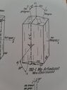
Very nice slide! You should also try to identify the pleochroism of the arfvedsonite. From your picture, one already recognizes a change from light blue to light green parallel or perpendicular to the longitudinal extension. How is your polarisatior oriented? (NS or EW)? Do you also find a typical "head-cut"? What are the pleochroitic colours there? The picture is from W. E. Tröger, Optische Bestimmung der gesteinsbildenden Minerale, Teil I Bestimmungstabellen, 5th edition, Schweitzerbartsche Verlagsbuchhandlung, Stuttgart, 1982. The book is an absolute must have!
Furthermore, I highly recommend a look at
https://homepage.rub.de/olaf.medenbach/download/mineraloptik/
although only in German.
 
Attachments
  • Crystal drawing of arfvedsonite, 183-1 Mg-Arfvedsonit, showing a prism with labelled faces 110, 101, 010, 011, axes a, b, c and angle marks 50 degrees and 65 degrees, with colour notes hell gelbgrün, hell blaugrün and fast opak.
    photo_5224555522487867383_y.jpg
    22.3 KB · Views: 136
Last edited:
DrDu said:
Very nice slide! You should also try to identify the pleochroism of the arfvedsonite. From your picture, one already recognizes a change from light blue to light green parallel or perpendicular to the longitudinal extension. How is your polarisatior oriented? (NS or EW)? Do you also find a typical "head-cut"? What are the pleochroitic colours there? The picture is from W. E. Tröger, Optische Bestimmung der gesteinsbildenden Minerale, Teil I Bestimmungstabellen, 5th edition, Schweitzerbartsche Verlagsbuchhandlung, Stuttgart, 1982. The book is an absolute must have!
Furthermore, I highly recommend a look at
https://homepage.rub.de/olaf.medenbach/download/mineraloptik/
although only in German.
Thanks for the feedback! I was busy last week working with my solar eclipse images, these are excellent questions/suggestions- thanks!
 
This week's sample is another trachyte:

Photo of a rock thin section mounted under a glass slide, label reading 'FEN 34g' in handwriting, showing a grey-blue mineral groundmass with pale rectangular and angular crystal fragments.


I chose this slide due to the presence of 'something' I'm hoping someone can help identify, that is also present in many other samples. I would describe this sample as having:

Phenocrysts of altered sanidine, altered nepheline, minor amounts of altered biotite in a groundmass of acicular aegirine, platy trachytic feldspar, and ‘granules’ of feldspar (?). Minor amounts of cabonatite. Nepheline phenocrysts are heavily altered to mica (?), almost appears like glimmerite, with a rim of granulated feldspar (?)

Here's a sequence of altered nepheline, showing the 'glimmerite' and granules of 'something' on the rim:

Photomicrograph in plane-polarised light of a trachyte thin section, with a 1 mm scale bar; a large altered grain sits at upper centre among a pale groundmass of fine acicular crystals and dark olive-brown patches.


Cross-polarised thin section photomicrograph at 1 mm scale of a trachyte: a large altered phenocryst with granular rim sits in a fine acicular groundmass, with small yellow-green grains nearby.


Cross-polarised light photomicrograph of a thin section showing a band of fine, granular crystals with bright interference colours, flanked by elongated yellowish-brown altered crystals and dark, nearly isotropic material. Scale bar reads 100 µm.


Photomicrograph in plane-polarised light of a fine-grained rock groundmass with interlocking elongated and acicular crystals; small dark opaque grains are scattered throughout. A 10 µm scale bar is at the lower right.


Thin section photomicrograph under cross-polarised light showing acicular, elongated crystals in yellow, orange and grey interference colours, with dark opaque areas and a 10 µm scale bar at lower right.


Photomicrograph in plane-polarised light of a thin section with a pale, clouded groundmass, scattered black opaque grains, and dark-brown altered crystal fragments; 10 µm scale bar at lower right.


Cross-polarised light photomicrograph of a thin section with a 10 µm scale bar; a mosaic of small bright granular crystals with dark patches and elongated colourful crystals at lower right.


Whatever that foamy stuff is, it's also present in the (altered) sanidine:

Cross-polarised thin section photomicrograph at 100 µm scale showing acicular crystals and clusters of granular, highly birefringent grains in a dark, altered groundmass.


The little bits of carbonatite are nicely photogenic- I identified these as carbonatite due to the high birefrengence- which also helps create a 3-D appearance:

Cross-polarised light micrograph of altered feldspar or nepheline grains in a thin section, with bright interference colours and a 20 µm scale bar in the lower right.
 
DrDu said:
Very nice slide! You should also try to identify the pleochroism of the arfvedsonite. From your picture, one already recognizes a change from light blue to light green parallel or perpendicular to the longitudinal extension. How is your polarisatior oriented? (NS or EW)? Do you also find a typical "head-cut"? What are the pleochroitic colours there?
I had a few images on hand to show pleochroism- here's a pair of PP and XP images with the sample rotated 90 degrees between the sets. On the upper pair of images, the arfvedsonite is along the left half and bottom half. The (according to my eyes) green-blue pleochroism is pretty cool :).

Four thin-section micrographs in a 2x2 grid: plane-polarised light on the left, crossed polars on the right, two sample orientations; irregular mineral grains with dark fibrous amphibole-like bands and vivid interference colours.


About your other questions- the polarizer is oriented (I think) NS, but that's a guess based on the angle marking on the polarizer mount (zero degrees). What does "head-cut" mean- a slice perpendicular to the long axis? There are a few square-ish/diamond shapes that have a strong green -> pink color change....? I can try and get some decent images of those.
 
DrDu said:
The white rim looks like quartz to me.
I was wondering if that's the case. Is it an alteration product? My understanding is that these rocks are all very silica-undersaturated.
 
Andy Resnick said:
I had a few images on hand to show pleochroism- here's a pair of PP and XP images with the sample rotated 90 degrees between the sets. On the upper pair of images, the arfvedsonite is along the left half and bottom half. The (according to my eyes) green-blue pleochroism is pretty cool :).

View attachment 343673

About your other questions- the polarizer is oriented (I think) NS, but that's a guess based on the angle marking on the polarizer mount (zero degrees). What does "head-cut" mean- a slice perpendicular to the long axis? There are a few square-ish/diamond shapes that have a strong green -> pink color change....? I can try and get some decent images of those.
The orientation of the polariser is best detected by the pleochroism of biotite. Biotite, cut perpendicular to the sheets, appears dark if the polariser is oriented parallel to the sheets, and bright if perpendicular.
And yes, a "head cut" (my translation of the german "Kopfschnitt") is a slice perpendicular to the long axis. You should see the characteristic cleavage pattern with 60 and 120 degree angles and also pleochroism, which is different, at least in one direction, from the colours seen along the longitudinally oriented crystals.
 
  • Like
Likes   Reactions: Andy Resnick
DrDu said:
The orientation of the polariser is best detected by the pleochroism of biotite. Biotite, cut perpendicular to the sheets, appears dark if the polariser is oriented parallel to the sheets, and bright if perpendicular.
And yes, a "head cut" (my translation of the german "Kopfschnitt") is a slice perpendicular to the long axis. You should see the characteristic cleavage pattern with 60 and 120 degree angles and also pleochroism, which is different, at least in one direction, from the colours seen along the longitudinally oriented crystals.
Last night I realized I need to be more careful in my polarizer description/analysis. The polarizer may indeed be oriented NS, but then the analyzer is EW, so for PP imaging I may need to be more specific about which polarizing element is in the path... stay tuned!
 
Andy Resnick said:
Last night I realized I need to be more careful in my polarizer description/analysis. The polarizer may indeed be oriented NS, but then the analyzer is EW, so for PP imaging I may need to be more specific about which polarizing element is in the path... stay tuned!
Usually, when talking about PPL, you assume that the polarizer is present, but the analyzer has been removed.
 
DrDu said:
The orientation of the polariser is best detected by the pleochroism of biotite. Biotite, cut perpendicular to the sheets, appears dark if the polariser is oriented parallel to the sheets, and bright if perpendicular.
And yes, a "head cut" (my translation of the german "Kopfschnitt") is a slice perpendicular to the long axis. You should see the characteristic cleavage pattern with 60 and 120 degree angles and also pleochroism, which is different, at least in one direction, from the colours seen along the longitudinally oriented crystals.
I had a chance to take a few careful images of arfvedsonite crystals today, I think I found an arrangement with both 'head cut' and longitudinally oriented crystals in the field of view. Here's a photo with just the (EW) analyzer present:

Photomicrograph in plane-polarized light of a thin section showing an amphibole crystal with fine cleavage traces, inclusions and a pale blue-gray region at right.


On the right side, the crystal is in the 'head-cut' position (I think, based on the fracture pattern), while at the lower left (and also a small one nestled in on the right) are longitudinally-oriented crystals. The same field of view, this time only the (NS) polarizer in place:

Photomicrograph of a thin section in plane-polarized light showing pale, colourless to greenish mineral grains with dark fractures and cleavage traces, with dark green-black grains scattered throughout.


I tried to orient the sample to maximize the color changes. Here's both analyzer and polarizer (crossed polars):

Photomicrograph in cross-polarized light of a thin section of arfvedsonite, showing blue, orange and purple interference colours across crystal grains with fracture lines.


And a montage of PP(both polarizer and analyzer) and XP under sample rotation:

Grid of eight microscope photographs of arfvedsonite crystals in thin section under varied polarized light, plus one black frame; the top row and two middle images are plane-polarized and the bottom-right images are cross-polarized with blue and yellow interference colours.


In the XP series, I measured an extinction angle of 25 degrees... assuming I did that correctly.

Then I just had some fun, playing with color: here's XP plus 1/4 waveplate:

Photomicrograph of a thin section under crossed polarizers showing brightly coloured interference colours in blue, purple, yellow and orange across elongated mineral grains.


Replacing the polarizer with a circular polarizer (circularly polarized light incident on sample):

Photomicrograph in cross-polarized light of a thin section showing interference colours of blue, yellow, purple and magenta across crystal grains with fracture lines.


And then 2 photos using only a Cokin chromofilter SA: I forgot to pay attention to the exposure setting, both of these are overexposed.

Photomicrograph of a thin section of arfvedsonite, with pink and magenta tones and bluish patches, showing fractured crystal grains under polarized light.


Photomicrograph in cross-polarized light of a thin section showing a pink to magenta field with mineral grains of varied relief, cleavage traces and dark inclusions.
 
This is a sample of an ultramafic rock:

Thin section of an ultramafic rock under cross-polarised light on a dark slide, with a handwritten label reading "69 Fen 50"; densely packed mineral grains show bright blue, pink and orange interference colours.


I think this is either a phlogopite-hornblende-olivine peridotite or a pyroxene peridotite- I had (and still have) trouble distinguishing between pyroxene and amphibole in this sample. Major minerals: serpentinized olivine phenocrysts, horneblende or clinopyroxene phenocrysts, some zoned/twinned. Phlogopite phenocrysts. Opaques may be chromite? Not much of a groundmass, but what there is consists of small grains of phlogopite and carbonatite.

Here's a pair of images showing phlogopite, calcite, what I think is apatite at bottom. The grain in the upper right corner is unknown (to me):

Side-by-side thin section micrographs under plane-polarised light (left) and crossed polars (right) showing elongated pleochroic yellow-brown crystals and an interference-coloured grain, with a 100 µm scale bar.


An oddly zoned phlogopite:

Plane-polarised light photomicrograph of a thin section showing a zoned yellow-brown crystal with a pale green core, with dark opaque grains and a 100 µm scale bar at lower right.


Here are a couple image pairs of what has confused me- grains that are either pyroxene or amphibole grains, I can't identify which:

Side-by-side thin section photomicrographs, plane-polarized light on the left and crossed polars on the right, with a 100 µm scale bar; a dark opaque grain with a tail sits among a fine, cross-hatched brownish-green mineral matrix.


Side-by-side thin section micrographs under plane-polarised (left) and cross-polarised (right) light, with a 100 µm scale bar; dark opaque grains and crystal fragments with fibrous, fractured textures.


Any guesses?

Here's another grain with some (relic?) serpentized olivine:

Side-by-side thin section micrographs in plane-polarized light (left) and crossed polars (right) of an ultramafic rock with elongated, rounded grains, scale bar 100 µm.


Finally, while I've posted a few images previously of the serpentinized olivine, it's so photogenic I decided to post some more images here again:

Photomicrograph in crossed polarised light of a thin rock section: a blue-banded vein with dark opaque inclusions runs across the top, crossing a bright orange-red vein that descends vertically at centre; a 100 µm scale bar is at lower right.

(XP with full wave plate)

Thin section photomicrograph under crossed polars showing a red and blue interlocking network of serpentinized olivine veins, with a 1 mm scale bar at lower right.


(Chromofilter SA)

Transmitted light micrograph of a thin section showing several dark, angular mineral grains on a grey background with pale diagonal banding and a 10 µm scale bar.


(XP)
 
This sample is very heterogeneous with some interesting and unusual textures:
Thin section photographed between crossed polarisers on a dark background; a heterogeneous grain with a bright blue crystalline mosaic on the left, dark granular and opaque areas in the centre, and a speckled, coloured porphyritic region on the right.


On the left side (side closest to label) consists of approximately equal amounts plagioclase (albite?), lightly sericitized K-feldspar (orthoclase?) and little to no quartz, so I classify that part (presumably country rock) as a monzonite with aegirine inclusions:

Photomicrograph in cross-polarised light of a thin section showing interlocking grains with bright lamellar twinning and dark opaque patches, with a 1 mm scale bar at lower right.


Right side is magmatic with a porphyritic texture: phenocrysts of pyroxene and hornblende, each with reaction coronas, opaques, and minor apatite in a groundmass of anhedral carbonatite and masses of skeletal phlogopite.

Photomicrograph in plane-polarised light of a heterogeneous thin section with yellowish-green pleochroic grains, clear feldspar-like grains, and a dark central grain; scale bar reads 100 µm.


Cross-polarized thin section micrograph at high magnification showing a heterogeneous mineral assemblage with bright interference colors, with a 100 μm scale bar in the lower right.


I'm not sure what the central grain is; it could be acmite (a fibrous form of aegirine)?

Reaction coronas (here, around a grain of hornblende) appear to consist of phlogopite pseudomorph surrounded by a shell of granular carbonatite and plagioclase, itself surrounded by a shell of skeletal phlogopite.
Thin section photomicrograph in plane-polarised light, scale bar 100 µm, showing brownish-yellow and orange mineral grains with colourless patches and fibrous pink-tinted prismatic crystals.


Photomicrograph in cross-polarised light of a thin section showing a mosaic of brightly coloured interference-coloured mineral grains, pale grey grains, and a large yellow-brown grain with fine parallel striations; scale bar 100 µm.


In between the two phases (magmatic and monzonite) is a transition phase consisting of massed prismatic and acicular aergirine, especially concentrated around macrocysts of monzonite.

Cross-polarised thin section micrograph under crossed polars showing bright interference colours on elongated and prismatic mineral grains against a dark grey background, with a 100 µm scale bar at lower right.


Photomicrograph in plane-polarized light of a thin section with a 1 mm scale bar, showing a light feldspar-rich matrix with dark opaque masses and clusters of thin dark acicular crystals.


Thin section photomicrograph under cross-polarized light showing a large, pale feldspar-like crystal with dark acicular needles, bordered by dark brown and black material; scale bar reads 1 mm.


How did the aegirine needles form inside the pre-existing feldspar crystals? Also, since the 'monzonite' part of my sample is only about a square centimeter, what confidence do I have that it is a representative sample of the country rock? (not much, but I'm no expert).

Some opaques in the magmatic part also appear to have a dendritic or skeletal texture. Flakes of 2 different Iron compounds (likely pyrite and magnetite) occur together- this image is reflected darkfield (in transmitted brightfield the whole frame would be black):

Photomicrograph in plane-polarised light of a thin section: pale fibrous and acicular grains fill most of the field, with a cluster of gold-brown opaque grains and a small blue-tinted acicular crystal near the centre. Scale bar reads 100 µm.


My suspicion is that the white mineral is aegirine, based on what other parts of the sample look like in reflected light.
 
This sample is part of a group all labeled 'Fen 6':

Cross-polarised thin section photomicrograph of a heterogeneous rock: a fine-grained granular mosaic on the left, a mottled grainy zone in the centre, and a darker breccia with angular fragments on the right; a label reads Fen 6 (NW 8D).


This heterogeneous rock is a carbonatite. On the left is crystallized carbonatite (probably calcite) with trace amounts of apatite. On the right is a carbonatized magmatic breccia, possibly a (carbonatized) damtjernite-like explosion breccia. In between is a narrow transition zone, likely re-melted and re-crystallized carbonatite; evidence of melting seen from some crystalline carbonatite grains at the boundary:

Thin section photomicrograph under cross-polarised light of a carbonatite showing granular calcite-like grains with bright interference colours, a pale irregular patch extending from top to bottom, and a 1 mm scale bar.


Cross-polarized light photomicrograph of a thin section showing carbonatite, with bright interference-coloured calcite grains in pink, blue and yellow against a dark, fine-grained matrix; scale bar reads 1 mm.


Different fields of view were used for PP and XP to highlight the selected, deformed, grain.

The transition zone itself consists of granular carbonatite and some apatite. The granular carbonatite looks like soap bubbles at high magnification:

Photomicrograph in plane-polarised light of a carbonatite thin section, showing a granular mosaic of pale calcite-like grains with dark rims, a scale bar reading 100 µm, and a smoother pale grain area at right.


Cross-polarised light micrograph of a thin section of granular carbonatite with a mottled, multicoloured interference pattern, scattered opaque grains and a few bright birefringent crystals; scale bar 100 µm.


Cross-polarised micrograph of granular carbonatite showing a mosaic of small, rounded grains with bright interference colours and dark boundaries, resembling soap bubbles; a few dark opaque grains and a scale bar of 10 µm at lower right.


The interface between re-melted carbonatite (upper part of image, below) and the breccia is marked by the appearance of opaques:

Thin section photomicrograph in plane-polarised light showing a light-coloured carbonate groundmass with dark granular opaque material and a single elongated, pale yellow-brown grain with internal striations, scale bar 1 mm.

Breccia consists of K-feldspar (some altered) and phlogopite phenocrysts in a groundmass of granular carbonatite, apatite, and opaques. The large altered xenocryst has a large equant massed phlogopite corona:

Polarized light photomicrograph of a thin section showing densely packed, interlocking carbonate grains in yellow, orange and grey with bright interference colours; scale bar reads 100 µm.


While many of the phlogopite phenocrysts have severe dislocations:

Thin-section photomicrograph under plane polarised light showing a carbonatite with a 100 µm scale bar; pale elongated grains are separated by a brown, layered vein-like zone running diagonally through the image.
 
This sample is a myrmekite:

Thin section photomicrograph under crossed polarisers showing a mosaic of blue, grey and yellow-orange mineral grains with a fine intergrown texture, mounted on a labelled slide reading 3 and Fi.


The large anhedral grains are pyroxene and K-feldspar; what appears to be a groundmass is the vermicular intergrowth of quartz and feldspar:

Photomicrograph in cross-polarised light of a myrmekite texture: vermicular, worm-like intergrowths of quartz and feldspar in a dark-blue and white groundmass, with large yellow-orange grains at the left and right edges. Scale bar reads 1 mm.


Cross-polarised thin section micrograph at high magnification showing a vermicular intergrowth of quartz and feldspar with larger grains of pyroxene and feldspar, scale bar 100 µm.


Photomicrograph in plane-polarised light of a thin section showing a large grey anhedral grain surrounded by a bright, vermicular intergrowth of quartz and feldspar, with a 100 µm scale bar.


Cross-polarised photomicrograph of myrmekite, showing large grains surrounded by a fine vermicular intergrowth of quartz and feldspar; scale bar 100 µm.


Getting decent high-magnification images of the myrmekite was, in places, challenging because the vermicular texture can be highly three-dimensional. Optically, that translates to an optical thickness (proportional to the integrated refractive index n(x,y)= ∫n(x,y,z)dz) having large spatial variations. In XP, this creates a photogenic effect:

Photomicrograph in plane-polarised light of myrmekite, showing vermicular intergrowths of dark worm-like quartz and feldspar threads on a pale background; scale bar 10 µm.


But PP imaging worked better using a low-NA lens and (digitally) zooming in for the desired magnification:

Photomicrograph in plane-polarised light of myrmekite, showing a dense vermicular intergrowth of worm-like grains against larger pale grains, with a 0.1 mm scale bar at lower right.


Cross-polarised photomicrograph of a thin section showing vermicular intergrowth of quartz and feldspar in a fine groundmass, with larger blocky grains at right; scale bar reads 0.1 mm.


In a few places I could really zoom in:

Photomicrograph in plane-polarised light of a thin section showing a vermicular intergrowth of quartz and feldspar with irregular grain boundaries, scale bar 10 µm.


Plane-polarised light photomicrograph of a thin section showing a large pale grain with dark, irregular curved boundaries and small dark inclusions; scale bar reads 10 µm.
 
This is another sample that was included in a publication:

Photomicrograph in cross-polarised light of a thin section with a mosaic of blue-grey and pale blue interference-colour mineral grains, with a label reading 69 Fen 33 on the glass slide edge.


Unlike many of the other samples in that publication, I have 8 samples identified as "69 Fen 33", 5 of which are not covered- the finished rock surface is exposed to air. I'm sure what to do about those- they appear very different without the canada balsam to sort-of index match, but I don't want to damage the samples by mishandling them or mounting them poorly. All 8 have slightly different compositions, so it's not clear which one corresponds to the actual published data:

Sample is a weakly fenitized gneiss: 20% quartz, 25% perthite, 35% plagioclase, 10%-0% anorthite. 10% biotite, 5% hornblende, accessory amounts of opaques, apatite, zircon, and allanite. Fenitization-1 product is 1% aegirine, Fenitization-2 products are 1% arfedsonite, 1% stilpnomelane and 1% carbonatite (in veins).

Oops- I hust realized I forgot to add scale bars... most of these images were shot with a 16x objective, FWIW.

I could locate grains of apatite, but regarding zircon- I don' think there is any in this particular sample (Fen 33 ii), but it does appear to have high-relief grains that fall on the clinozoisite <—> epidote axis: clinozoisite is colorless, epidote is ‘fluorescent’:

Photomicrograph in cross-polarised light of a thin section: a cube-like colourless crystal with high relief and internal fractures sits in a blue-grey mineral matrix, with a dark brown-black grain below and a yellow birefringent grain at lower left.


Photomicrograph in plane-polarised light of a thin section: a dark brown elongated mineral grain with two rounded, speckled grains inside it, set against a pale yellowish-brown background and a white area.


Photomicrograph in cross-polarised light of a thin section showing a brown mineral grain with fine cleavage lines, containing two small high-relief grains with bright interference colours, surrounded by pale grey and dark mineral areas.


Zircon also has very high relief, so perhaps these are indeed zircon.

I think this is a grain of allanite (because of the radial fracturing):

Thin section photomicrograph under cross-polarised light showing a small oval yellow-green grain with a dark outline, surrounded by grey-blue mineral grains, with brownish-yellow grains at the right and bottom edges.


But there isn't any halo of radiation damage (I couldn't find any such halos in any of the Fen 33 samples), so...?

Many of the biotite grains are either highly degraded or have a fine-grained corona when in contact with quartz, no corona is present when in contact with feldspar and precipitated opaques. I really like this example because if you look carefully, the plagioclase twinning is visible 'through' the corona:

Photomicrograph in plane-polarised light of a thin section showing an elongated yellowish-brown mineral grain with dark opaque inclusions, surrounded by pale, granular material and a greenish-brown grain below.


Photomicrograph in cross-polarised light of a thin section of rock showing a large yellow-brown mineral grain with dark opaque inclusions, beside a grey mineral with fine parallel cleavage or twin lines and a smaller yellow-green grain.


Not sure how that happens...

What I assume are altered biotite flakes have been replaced with 'stuff', the only thing I can positively identify is stilpnomelane (finally, I spelled it without having to look it up!)- it's the furry ring surrounding a grain of quartz:

Photomicrograph in plane-polarised light of a thin section showing a rounded brown-rimmed grain surrounded by pale mineral grains and dark opaque specks.


In some places, the quartz grains are loaded with minute inclusions: some are spherical (gas? liquid?), many more are not. And there's this chunk- no obvious fractures or twinning present, but there are hints of 120/60 degree axes and apparent zoning (unfortunately, hinting at 90 degree axes), so maybe this is hornblende:

Thin section photomicrograph in plane-polarised light showing a brown, finely speckled rock matrix with scattered dark opaque grains and thin dark needle-like crystals.


Photomicrograph in plane-polarised light of a thin section of weakly fenitized gneiss, showing a mottled mineral matrix with many small dark opaque grains and a faint pale grain outline near the centre.


Ideas on if/how to handle uncovered specimens are welcome, as are suggestions on the unidentified minerals!
 
If it helps, here's a pair of images from a different Fen 33 sample showing grains of apatite (ovoid, top left), altered biotite (top right), perthite (center), zircon/clinozoisite (center and lower left), and hornblende (?) (left and bottom):

Photomicrograph in plane-polarised light of a thin rock section showing pale colourless grains, dark green and brown altered mineral grains, and small black opaque inclusions.


Cross-polarised light photomicrograph of a thin rock section showing interlocking mineral grains in grey, orange, brown and dark green with a dark crack running through the centre.


In the very center is a crack/fracture in the sample (black in XP).
 
This sample is a weakly fenitized carbonatite:

Thin section photomicrograph under crossed polars showing a mottled, granular mosaic of pale grey and bluish carbonate grains, with scattered dark opaque specks; label on the slide reads "MA 72, 600 m".


Most of the fenitized samples I have were originally telemark gneiss (the country rock), so this one is of particular interest, as it demonstrates some chronological ordering between the carbonatite intrusion event and the fenitiztion event. According to the relevant paper, there were 2 fenitization events; the first one was a high temperature dehydration reaction while the second was a low temperature hydration and carbonation event- both are types of contact metamorphic events. This sample emplaces the carbonatite intrusion after the 1st fenitization but before the 2nd. Unsurprisingly, most of the sample is carbonatite (90%), but there is a reasonable amount of apatite as well (5%), in its characteristic anhedral, vaguely glomerular, habit.

The metamorphism is seen when examining biotite grains; these are surrounded by a corona of massed anhedral carbonatite grains:

Thin section photomicrograph in plane-polarised light at 1 mm scale bar, showing a dark biotite grain surrounded by pale carbonate grains, with a bright yellowish birefringent mineral grain at its centre.


Cross-polarised thin section photomicrograph of carbonatite with scattered black opaque grains, pale calcite, and a bright interference-coloured grain at centre; scale bar 1 mm.


Photomicrograph in plane-polarised light of a thin section showing pale anhedral carbonate grains with cleavage traces, surrounded by a dark, granular fine-grained alteration zone. Scale bar reads 100 µm.


Photomicrograph in cross-polarised light of a thin section showing a bright, multicoloured interference-coloured grain at left, a dark isotropic-looking grain at right, and a grey-green matrix; scale bar reads 100 µm.


Photomicrograph in plane-polarised light showing a grainy, pale carbonate matrix on the right and a finer, streaky textured area on the left, with a scale bar reading 10 µm in the lower right.


Polarised light photomicrograph of a thin section: a birefringent, colourful grain mass on the left grades into a pale, granular carbonate-rich area on the right; 10 µm scale bar in the lower right.


In several places, the biotite has been replaced by chlorite and quartz:

Photomicrograph in plane-polarised light of a thin section showing pale carbonate grains with green-brown chlorite-like patches and dark opaque specks; scale bar reads 100 µm.


Thin section photomicrograph in cross-polarised light at 100 µm scale, showing pale grey-white carbonate grains with dark, speckled mineral aggregates along the edges, including dark blades and a larger tan background region.


And located within the carbonate (I suspect it's all calcite) are random cabonatite grains that have a relatively lower birefringence so they stand out from the crowd:

Photomicrograph in cross-polarised light of a thin section showing a mosaic of anhedral grains in dark, grey and bright interference colours, with a 1 mm scale bar at lower right.


According to the paper, this sample is 90% (primary) carbonatite, 1% biotite, 5% apatite, 1% opaques, and the low-temp hydration-carbonation metamorphism produced 3% chlorite and accessory amounts of rutile, stilpnomelane, quartz, and fenitized carbonatite; the remarks state "
WF2carbonatite; F2Chl,F2Qtz,Stp veins; Bt⇒F2Chl,F2Qtz,F2Carb rims/veins"

I didn't see any rutile or stilpnomelane in this section, and I also wonder what a carbonated carbonatite would look like?
 
Thin section photomicrograph under cross-polarised light, with a paper label reading FEN 283 along the left edge; a light-coloured banded band at the top above a dark, grainy mineral mass with bright interference colours.


I chose this sample, possibly showing the chilled margin of an intrusion into country rock, because of the high concentration of sphene (a titanium-bearing mineral):

Cross-polarised thin section photomicrograph with a 100 µm scale bar showing a large pale brown sphene crystal surrounded by smaller colourful mineral grains and a coarser grey-green matrix.


Cross-polarised light photomicrograph of a thin section showing a mosaic of mineral grains with bright interference colours, including pinkish and yellow-orange grains, grey-blue grains and dark opaque areas. Scale bar reads 100 µm.


The magmatic component of this sample has a relatively high concentration of sphene (more than 10%) in a matrix of carbonatite, apatite, pyroxene (augite?) and heavily sericitized orthoclase. A small vein and smaller veinlet, both full of calcite, pass through the magmatic portion. Here's a pair of images of the veinlet as it passed through sericitized orthoclase:

Transmitted light photomicrograph of a thin section showing a diagonal calcite vein crossing a fine-grained mineral matrix, with a scale bar of 100 µm at lower right.


Photomicrograph in cross-polarised light of a thin section showing a pale calcite veinlet running diagonally through a mottled matrix of colourful birefringent grains; scale bar reads 100 µm.


The calcite veinlet is surrounded by some intermediate mineral, but I can't identify what it is.

The country rock (Telemark gneiss) has been strongly altered into perthite and chessboard albite:

Cross-polarized photomicrograph of a thin section of rock at 1 mm scale, showing grey and dark interlocking mineral grains with irregular, sutured boundaries.


Of interest (to me) are the sutured/interlobulated grain boundaries.

Cross-polarised light photomicrograph of a thin rock section with a 1 mm scale bar, showing interlocking mineral grains in bright blue, yellow and grey tints, with dark, altered patches and a thin crack running through them.


in the above, the lower left portion is sericitized orthoclase. I believe the light blue and yellow tints are evidence that the thin section is slightly thicker than 30 microns, but alternatively I may have inserted a 1/4λ plate. In any case, another interesting feature of this sample are the sphene grains near the margin boundary; the metals appear to have precipitated, turning the grains opaque:

Cross-polarised light photomicrograph of a thin section with a 100 µm scale bar, showing densely packed, brightly coloured mineral grains with several large opaque black grains in the centre-right.


In the above, it's clear that the group of sphene crystalline grains have become opaque (one is partially opaque). Looking at one in reflection shows possible phase separation of the metal oxides:

Photomicrograph of a thin section in reflected light showing an irregular dark, granular grey-black patch with pale white crystalline material around its edges, on a blue-grey background; scale bar of 100 µm at lower right.


My first thought is that the dark grey corresponds to an iron oxide (siderite? magnetite?) while the light colored grains are titanium dioxide. But it's not clear that's the case, as the dark grey could also be ilmenite.

Ilmenite is supposed to be highly bireflective, but I'm not sure I can perform that technique. Epi-darkfield does not preserve the polarization state, and epi-brightfield was difficult due to reflection off the coverglass. I was able to to cancel out reflection from the coverglass by using crossed polarizers, but I think enough light passed through the sample, reflected off the bottom of the glass slide, and then passed back through the sample so that I was essentially observing transmitted birefringence.
 
This sample is prominently featured in: https://www.ngu.no/filearchive/NGUPublikasjoner/NGUnr_380_Bulletin_70_Verschure_35_49.pdf.

Thin section photomicrograph under cross-polarised light of a breccia with a fine-grained grey groundmass containing many rounded and angular xenoliths; a large bluish-grey crystal cluster occupies the upper left, and a label reads 75-HTJ 5.


As presented in the paper, this sample originated in Hönstjern (UTM coordinates 5354-65418) and is a carbonatized damtjernite-like explosion breccia. Using K-Ar dating on biotite, this sample was assessed resulting in a calculated age of 578 Ma. This sample is one of two given considerable text in the paper. Quoting the paper (I will insert my images here and there:

“Two explosion breccias produce ambiguous and conflicting age data:

In the Bamble region two carbonatized damtjernite-like explosion breccias have been studied, the Hönstjern breccia and the Tveiten breccia. They are situated less than 0.5 km apart, in an area dominated by anatectic paragneisses with intercalated amphibolites and metagabbros (Morton et al. 1970). The breccias lie about 10 km W of the nearest exposure of Permian intrusives of the Oslo Graben. The breccias are very similar; they consist of a wide variety of xenoliths and xenocrysts in a very fine-grained groundmass consisting mainly of carbonate, green biotite, opaques and apatite. Among the xenoliths three groups can be distinguished: (1) small, rounded fragments (up to 0.5 cm in diameter) of ultramafic, occasionally porphyritic rocks with phenocrysts of biotite or brown hornblende;

Cross-polarised thin section photomicrograph of a fine-grained carbonate-rich breccia with rounded and angular xenoliths of varying size, scale bar 1 mm.


(2) larger, angular fragments (up to 10 cm in diameter) of crustal gneisses, amphibolites, granites and metagabbros; and (3) occasional fragments of a similar damtjernitic breccia. Many of the xenoliths and xenocrysts are strongly altered, but the abundant apatite phenocrysts and the cores of biotite phenocrysts and perthite xenocrysts do not show any alteration.

Photomicrograph of a thin section in plane-polarised light showing a fine-grained dark groundmass with scattered pale, angular crystals and yellowish-brown flakes, with a 1 mm scale bar in the lower right.

[Note: transparent apatite and green-brown biotite phenocrysts in the dark-colored groundmass]

Cross-polarized thin section micrograph showing a sharp boundary between a region of radiating fibrous crystals at upper left and a fine-grained speckled groundmass with bright grains below; a scale bar reads 100 µm.

[Note: perthite along the top, what I think is quartz along the bottom, and carbonate veinlets. I thought perthite resulted from metamorphic processes and would be considered an alteration, as is the quartz (higher magnification below, XP]

Thin-section photomicrograph in blue-tinted transmitted light showing a fine-grained groundmass with dark, irregular outlines around pale patches, with a 10 µm scale bar at lower right.


Numerous veinlets of carbonate transect both the xenoliths and the groundmass.

Cross-polarized photomicrograph of a thin section showing a rounded, banded carbonate-rich fragment with interference colours surrounding a dark central void, set in a fine-grained dark groundmass; scale bar 100 µm.

[Note: on left is sericitized plagioclase , right is perthite, center is carbonate]

Biotite phenocrysts from the Hönstjern breccia yield a K-Ar age of 578± 20 Ma, concordant with the age of the Fen complex. The partly chloritized biotite booklets from the Tveitan breccia give much younger ages, however: a K-Ar age of 280±10 Ma and a Rb-Sr model age between about 310 Ma and 255 Ma, depending on the assumed initial 87Sr/86Sr ratio (0.702 and 0.705, respectively). The early Permian age of the Tveitan biotite is supported by four K-Ar whole-rock dates obtained from the same breccia: an age of 316 ± 10 Ma for the groundmass and ages between 500 and 380 Ma for three crustal xenoliths. The latter three ages could very well be interpreted as reflecting varying degrees of resetting of the K-Ar systems of Sveconorwegian crustal fragments during transport by the exploding magma in Permian time.

[Data for K-Ar dating is now presented. Fun fact: In 2013, the K–Ar method was used by the Mars Curiosity rover to date a rock on the Martian surface, the first time a rock has been dated from its mineral ingredients while situated on another planet… thanx, wiki! The paper continues…]

There thus appears to be a difference between the age of the Hönstjern breccia and that of the Tveitan breccia; about 580 Ma for the former and about 280 Ma for the latter. The simplest explanation is that the age difference is real, the Hönstjern breccia håving been formed in the latest Precambrian, in relation to the damtjernite volcanism elsewhere, and the Tveitan breccia having formed in the Permian and associated with the magmatism in the nearby Oslo Graben. The similarity between both breccias is then difficult to understand, however. Another explanation could be that both breccias were formed about 280 Ma ago, but that the Hönstjern breccia contains biotite derived from an older rock, carried upwards by the exploding magma. “

I’m not sure the age discrepancy was ever reconciled…. Also, note that the features described as “small, rounded fragments (up to 0.5 cm in diameter) of ultramafic, occasionally porphyritic rocks with phenocrysts of biotite or brown hornblende” are now thought to be formed by stages of fluidized granulation (https://www.nature.com/articles/ncomms1842), similar to a spray coating process.

A few remaining images: first, a grain of apatite that seems to have zoned inclusions of carbonate:

Side-by-side thin section photomicrographs at 100 µm scale, plain light on the left and crossed polars on the right, showing an elongated crystal with a dusty, inclusion-rich rim and internal cracks, surrounded by other grains in a fine matrix.


What I think is chlorite (and some quartz along the left and top edge; a small spear of carbonate radiating right and up):

Photomicrograph in plane-polarised light of a thin section showing a fine-grained brown-green groundmass with a central cluster of altered grains and radiating dark needle-like crystals, with pink-stained crystal fragments at the left and top edges. Scale bar: 100 µm.


Photomicrograph in cross-polarised light of a thin section showing a radiating spherulite-like aggregate of fine acicular crystals with a dark centre, and a bright birefringent grain at upper right; scale bar 100 µm.


And a tiny 'snowflake', imaged with epi-darkfield:

Photomicrograph of a thin section showing a hexagonal, pale grain surrounded by a ring-shaped halo, with a 30 µm scale bar in the lower right.


This sample really opened up my understanding of a dozen or so other samples, I'll continue presenting other samples of carbonatized explosion breccias from Tveitan and Fen (near Söve).
 
Thin section photomicrograph under crossed polarised light of a breccia, showing angular mineral fragments and rock clasts in a fine-grained groundmass, with a large dark opaque area at centre and a handwritten label reading 'TVE 2' on the left edge of the slide.


This sample is classified as a Tveitan carbonatized damtjernite-like explosion breccia. It was obtained from UTM coordinates 5356-65419, and was dated to 316 Ma. From the paper: "[The breccia consists of] a wide variety of xenoliths and xenocrysts in a very fine-grained groundmass consisting mainly of carbonate, green biotite, opaques, and apatite. [...] Many of the xenoliths and xenocrysts are strongly altered, but the abundant apatite phenocrysts and the cores of biotite phenocrysts and perthite xenocrysts do not show any alteration. Numerous veinlets of carbonate transect both the xenoliths and the groundmass”. This sample has several angular fragments of xenocrysts and small pelletal lapilli of ultramafic rocks with cores of biotite or brown hornblende. Recall that even though this sample was obtained a few hundred meters from the explosion breccia at Hönstjern and appears very similar, the age of the two samples is completely different.

I wanted to image the pelletal lapilli which posed a challenge due to both the ultramafic nature and complex microscopic structure of the fragments. For example, here are three, showing both PP and XP views:

Six thin-section micrographs in a 2x3 grid, plane-polarised light on the left and crossed polars on the right, showing pelletal lapilli with dark rims and altered cores in a fine-grained groundmass; scale bar 1 mm.


It's difficult to tell what is going on, even at moderate magnifications. Zooming in on one of the cores located within the topmost one:

Photomicrograph in plane-polarised light of a thin section showing a fine-grained yellow-green groundmass with elongated black opaque grains and a brown altered fragment; scale bar 100 µm.


Cross-polarised light photomicrograph of a thin section showing a fine-grained groundmass with elongated dark opaque grains and bright interference colours in yellow, blue, magenta and green; scale bar reads 100 µm.


Photomicrograph in plane-polarised light of a thin section showing elongated black opaque grains and streaks scattered through a fine-grained yellow-brown groundmass, with a 10 µm scale bar in the lower right.


Photomicrograph in cross-polarised light of a thin section showing dark elongated opaque grains against a fine-grained groundmass with bright interference colours; scale bar 10 µm.

this one has a core of biotite (which has been altered but is still micaceous) interspersed with curved branching opaques.

The microstructure can be better viewed with epi-darkfield (this is the middle row lapilli):
Photomicrograph of a thin section showing a dark olive-brown angular rock fragment with pale, speckled inclusions set in a grey groundmass; a white scale bar labelled 1 mm lies below.

Here, the biotite core is surrounded by a shell of what is now (I think) chlorite and decorated by small high-index granules.

These granules are not opaque, but have a very high index of refraction and birefringence, so I suspect these are a titanium oxide. Here are additional high-magnification views of these granules and matrix, in the XP view it's a little hard to tell what is in focus and what is not:

Photomicrograph in plane-polarised light of a thin section of carbonatized breccia, showing rounded and angular altered xenoliths in a fine-grained brownish groundmass with dark opaque grains and veinlets; scale bar 10 µm.


Photomicrograph in cross-polarised light of a thin section showing a fine-grained groundmass with bright interference colours, dark opaque patches and a scale bar of 10 µm.


Photomicrograph in plane-polarised light of a thin section showing bright white angular grains, likely carbonate or feldspar fragments, set in a dark brown fine-grained groundmass with a 10 µm scale bar at lower right.


What is surprising to me is the roughness of the granules at the microscale- I would not have expected that, even for rapidly cooled magma.
 
A very photogenic (I think) diorite from Tveitan:
Photomicrograph of a thin section under crossed polarised light showing densely packed, elongated grey and white crystals in a random, interlocking arrangement, with a handwritten label reading '5 TVE 6' on the left.


This sample consists almost completely of cumulate plagioclase (including chessboard albite) with apatite and stilpnomelane, muscovite and chlorite (?) after biotite (if that's the correct phrasing): I mean that the original biotite crystals have, to a greater or lesser degree, been metamorphosed into stilpnomelane, muscovite and (I think) chlorite. There are minor amounts of carbonate and diopside. Plagioclase is heavily altered (‘dusty’ appearance). Clusters of euhedral and subhedral apatite grains, sometimes with interstitial stilpnomelane, are present:

Side-by-side photomicrographs of a thin section: left in plane-polarized light showing brown altered biotite flakes among pale plagioclase, right in cross-polarized light with a 1 mm scale bar showing colourful birefringent crystals.


Side-by-side micrographs of a thin rock section at 1 mm scale: plane-polarised light on the left, cross-polarised on the right, showing a brown, birefringent altered mineral patch within a pale plagioclase grain.


Thin section photomicrograph in plane-polarised light showing brown-yellow fibrous mineral aggregates, possibly stilpnomelane, around pale grey and pinkish crystal grains, with dark opaque specks; 100 µm scale bar at lower right.


Cross-polarised light photomicrograph of a rock thin section showing orange and yellow fibrous radiating aggregates with greenish-brown granular material, surrounded by grey striated crystals; scale bar 100 µm.


There are regions of disorganized "something" as seen by very disorganized birefringence that occur near a carbonate particle: Lower left corner is stilpnomelane, center is a carbonate agglomeration.

Side-by-side thin section micrographs in plane-polarised light (left) and crossed polars (right), scale bar 100 µm; the right image shows a rounded, birefringent carbonate-like grain surrounded by dark altered material.


I suppose the material could be quartz but regardless of what it is, I would say the optical evidence appears to show immiscible fluids. I want to re-visit this region and take some better images to really visualize the interface.

Other photogenic features occur within some of the altered biotite:

Photomicrograph in plane-polarised light of a thin section showing small red and green needle-like and star-shaped crystals with a hexagonal outline on a yellow background, with a 20 µm scale bar.


Photomicrograph in cross-polarized light of a thin section showing a small bright blue rounded crystal with dark edges, surrounded by colorful birefringent acicular grains, with a 20 µm scale bar in the lower right.

Suspended within what I think is chlorite are microcrystals of jewel-toned diopside and acicular stilpnomelane, possibly some hexagonal apatite as well.

An otherwise great image of biotite reminding me of a 3-D surface, marred by a dirty sensor:

Photomicrograph in plane-polarised light of a thin section showing curved, closely spaced parallel striations in brown and tan tones, with a small dark curved fibre at the upper right.


Lastly, within a grain of apatite is a microinclusion (possibly diopside?) showing stress birefringence:

Side-by-side micrographs of a dark brown mineral grain with radiating cracks in pale matrix, left in plane light and right in cross-polarised light with a 10 µm scale bar.
 
This sample has a lot of interesting and photogenic features:

Thin section of a carbonatite rock under cross-polarised light on a dark background, showing a mosaic of colourful interference-coloured grains with a label reading 6 FEN 22.


This sample is a mixture of calciocarbonatite and altered phlogopite. Composition is subhedral equigranular carbonate (likely calciocarbonate) grains, aggregates of subhedral apatite, and zoned phlogopite (orange-tan pleochrosim) with equigranular aphanitic carbonate reaction rims (phlogopite often strongly altered).

Photomicrograph in plane-polarised light of a thin section showing blocky, cross-hatched carbonate grains with orange-brown altered mica, a dark opaque cluster, and a 1 mm scale bar at lower right.

Carbonatite grains (likely calciocarbonatite), altered phlogopite with carbonate rims, and radiating prismatic aggregates of an opaque. For that particular object at high magnification, what appears to be a prismatic habit is actually a 'cloud' of fine opaque particles, almost like a smear. Possibly an artifact of lapping/polishing? I have similar features on other samples.

Cross-polarised light photomicrograph of a thin section showing zoned phlogopite with vivid interference colours, blue-grey apatite grains and opaque minerals, with a 100 µm scale bar in the lower right.

Carbonatite, aggregate of apatite, and biotite with carbonate rims. Some opaques at upper center.

Photomicrograph in plane-polarised light of a thin section: orange-tan pleochroic zoned mica grains bordered by white carbonate, with black opaque grains on the left. Scale bar 1 mm.

Zoned phlogopite (center) eroded by carbonate. Apatite aggregate on left, altered phlogopite on upper left and right side, some opaques on left.

A close-up of the carbonate rims:

Polarised light micrograph of a thin section showing a pale, grainy carbonate vein cutting through strongly birefringent green, orange and red phlogopite; scale bar reads 100 µm.


Birefringence of (calcite?) is so high that interference occurs at twinning planes, resulting in visible colors.

Thin section photomicrograph in cross-polarised light of calcite grains showing bright interference colours along closely spaced twin lamellae, with a 1 mm scale bar.

Carbonatite in PP.

Minor amounts of radiating splays of highly birefringent prismatic crystals: because the thickness of individual prisms is less than 30 microns, I instead estimated a thickness (10 microns), indicating aragonite.

Cross-polarised thin section photomicrograph of a rock showing blue and yellow interference-coloured prismatic bundles and crystals among grey and blue grains, with black opaque grains; scale bar of 100 µm.


Carbonatite on left side and right side, aggregate of apatite in center, prismatic bundles of something from lower left towards upper center, and opaques scattered throughout. It may not be apparent in this shrunken image, but it's possible to see through the prisms to the apatite underneath. At high magnification a single prismatic crystal looks like this (PP and XP):

Two side-by-side photomicrographs of a thin section, plane-polarised on the left and cross-polarised on the right, showing a prismatic crystal with striped internal texture; the right panel has vivid blue and yellow interference colours. Scale bar reads 10 µm.


The altered micaceous mineral consists of equigranular aphanitic aggregates, similar in appearance to carbonate reaction rims, but yellow color, no pleochroism, and birefringent color consistent with members of the biotite group, so it is most likely phlogopite as well.

Side-by-side thin section micrographs at 100 µm scale: left in plane-polarised light, right under crossed polars showing bright interference colours in the carbonate grains and altered rim.

Top is carbonate, lower 2/3 of image is filled with a granular phlogopite-like mineral.

Minor amounts of opaques. Some opaques within the phlogopite exhibit what appear to be pleochroic halos…. I wonder if this rock is radioactive?

Side-by-side thin section micrographs, plane-polarised left and cross-polarised right, showing a dark opaque grain surrounded by a pale carbonate rim; scale bar 100 µm.


The catch-22 is that if I borrowed a Geiger counter and this sample is indeed radioactive, campus safety people would likely confiscate it or require that I store the materials as hazardous. I'll pursue "don't ask, don't tell", as it were....