Random Photos

  • Thread starter Point Conception
  • Start date
  • Tags Tags
    Photos Random
4,009 replies · 305K views · last reply
BillTre said:
Frida Kahlo
My wife & I were visiting Mexico City. We saw her house and took the tour without knowing anything about her beforehand. Very cool.
 
  • Like
Likes   Reactions: DennisN and BillTre
gmax137 said:
My wife & I were visiting Mexico City. We saw her house and took the tour without knowing anything about her beforehand. Very cool.
There is also a good movie about her life.
 
gmax137 said:
My wife & I were visiting Mexico City. We saw her house and took the tour without knowing anything about her beforehand.
I had issues parsing this, until I realized that you were replying to the post above yours. Doh! o0)
 
  • Like
  • Haha
Likes   Reactions: gmax137 and BillTre
BillTre said:
So that's where Frida Kahlo is from?
I think so. According to Frida (given name) (Wikipedia):

Wikipedia said:
Frida (with variants Freda, Frieda, Frinta and Freida) is a feminine given name. In central and eastern Europe, Frida is a short form of compound names containing the Germanic element fried meaning "peace".

And according to this Swedish wikipedia page (translated with Google translate):

Swedish Wikipedia said:
Frida is an Old Norse female name formed from the word fridh which means the beloved, the beautiful. It is also a short form of German names such as Frideborg, Alfrida, Elfrida and Valfrida. The oldest evidence in Sweden is from the year 1388.

Edit: Though I got a bit confused by the Swedish Wikipedia page saying "fridh" means "the beloved, the beautiful", because there is a Swedish word "frid" which means "peace" (or "calm"), which seems to correspond to the quote from the English wikipedia page above.
 
from CNN:

Paratrooper descending under a brown round parachute beneath a grey military transport aircraft in pale sky, with a caption reading: A French paratrooper parachutes from an aircraft during a large-scale military drill in Castres, France, on Saturday, February 25.
 
  • Wow
Likes   Reactions: DennisN and collinsmark
Here two of my half Japanese nephews in Philadelphia. The one on left just completed masters in electrical engineering at University of Pennsylvania. And already has job at Ford motor company in Michigan.
Two young men smiling outdoors at dusk beside a river and waterfront promenade, city buildings and street lamps behind them; the man on the right holds up a phone showing a red screen.
 
  • Like
Likes   Reactions: collinsmark, DennisN, BillTre and 1 other person
Tom.G said:
And the one on the right?
Freshman engineering/business major at University of Rochester.
 
After patiently waiting for 10+ years (!!!), a 35mm faceplate for a Zeiss Ultraphot II became available on ebay and I immediately grabbed it. I have an Ultraphot III, but the piece fits well enough to use. Here's a quick pic of the new setup, with a dummy camera mounted where my Nikon camera goes:

Photo of a Zeiss Ultraphot microscope with a camera mounted on its top housing, a dummy camera body fitted to the 35mm faceplate, and a black specimen stage on a cluttered lab bench.


Before, my use of a camera on the Ultraphot was somewhat restricted- I could "only" use the extended optical path (designed for 4x5 film) like this:

Photo of a Zeiss Ultraphot microscope with a dark camera housing mounted on its top, set on a lab bench beside bottles and papers.

I'm not complaining- the image quality is fantastic- but I could only image using epi-brightfield. And, while I have the 4x5 film holder, medium-format film is not a road I am ready to travel down.

Photography using the 'normal' imaging path is restrictive because the image circle is about 22mm (C-mount specification), resulting in images looking like this (8x epi-darkfield):

Microscope view of a silicon chip corner showing rows of metal interconnect traces, a red-tinted region, and small square bond pads, with a wire bond visible below.


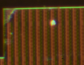
It doesn't look terrible unless you look closely: off-axis the image degradation (chromatic aberration, field curvature, etc.) happens fast- here's a 1:1 crop just off of center:

Microscope photo at high magnification of a dark, dense grid of vertical reddish-orange stripes on a black background, with a bright green-yellow spot near the top center and a glowing bright edge along the top and left border.

Using the 35mm film adapter, the image circle is magnified to better fill the sensor:

Microscope photograph of a silicon chip die showing a large rectangular block with fine parallel lines and a central square structure. Printed mirrored text on the block reads "2312" and "KSCCCCBGUC", with a small circular mark.


And while there is now some distortion and field curvature (which I think I can correct), the off-axis image characteristics are much improved: note that this crop has been downscaled about 60% to approximate the field of view of the prior crop:

Microscope photo of a dark-field image of a semiconductor chip showing repeating vertical bright-orange stripes with a bright yellow spot near the top center.


However, the real leap forward is not the image quality, it access to different imaging techniques- techniques that I could not use for photography because the luminar head doesn't support techniques like darkfield, polarization, DIC, phase contrast.... but now I can! Here's a epi-DIC image (downscaled to 800 pixels wide) using a 40x lens:

Microscope photo of an integrated circuit die showing an octagonal inductor coil of many concentric metal traces, with a bond pad array and striped wiring nearby, taken with reflected light.


I feel like I unlocked a whole bunch of new features! In practical terms, this means I have about 20 lenses that I can now use to give me (hopefully) amazing photographs. Currently, the only significant non-fluorescent imaging modality I can't do is epi-phase, and I am close to being able to implement that- I'm curious what epi-phase images look like since I have never seen any, anywhere.
 
Attachments
  • Micrograph of a regular grid of vertical reddish-brown stripes with green-tinted edges and a bright green-yellow spot near the upper right, with a green border along the top and left, likely a microscope image of a patterned surface.
    1678210596316.png
    29.5 KB · Views: 138
  • Like
  • Wow
Likes   Reactions: collinsmark, Rive, DennisN and 3 others
Nice picture of Portland's Japanese garden (not taken by me):

Moss-covered, gnarled tree with bare twisting branches in the foreground, misty garden with a small arched footbridge over a pond in the background, green shrubs at right.
 
  • Like
  • Wow
  • Love
Likes   Reactions: pinball1970, Klystron, collinsmark and 3 others
One small step for a yeti...

Close-up photo from above of a dark leather boot stepping into deep white snow, with snow dusted over the dark trouser leg.


...one giant leap for yetikind.

Close-up photo of a single boot print pressed into fresh snow, showing a lugged sole pattern with a star-shaped tread design.


We've got a bit of snow storm in the city... it was fun to be out and shooting, and I was happy that my camera is weather sealed, because this time it sure was needed :smile:.

Night street scene in heavy snowfall: a dark car with its headlights on and a taxi roof sign drives toward the camera over snow-covered road, with parked white vans and a red car along the left curb and blowing snow in the air.
 
  • Like
Likes   Reactions: collinsmark, Jonathan Scott, Borg and 1 other person
A piece of ice:

Close-up photo of a thick, irregular chunk of clear ice lit from behind, with trapped air bubbles, fractured edges and faint iridescent blue and purple tints against a dark blurred background.
 
  • Like
  • Wow
Likes   Reactions: collinsmark, Point Conception, BillTre and 4 others
Self-portrait (I licked a slide):

Phase contrast micrograph of a single epithelial cell, with a central oval nucleus surrounded by granular cytoplasm and a faint outline of the cell membrane.


100x phase contrast oil objective (transillumination). It's an epithelial cell, the center oval is the nucleus. The slightly out-of-focus lines are crosshairs from a focusing aid buried deep inside the camera adapter.
 
  • Like
Likes   Reactions: collinsmark, BillTre and DennisN
Andy Resnick said:
Self-portrait (I licked a slide):
So it is a cellfie? :smile:
 
  • Haha
  • Like
Likes   Reactions: collinsmark, Tom.G, BillTre and 5 others
Two tulips in May last year:

Two red tulips on slender green stems in sunlight, set against dark shadowed foliage with a tree trunk at left and white-flowered ground cover below.


(Lens used: my cheap but very nice Konica 40mm f/1.8)
 
  • Like
Likes   Reactions: Klystron, collinsmark, berkeman and 1 other person
A beautiful photo of a squirrel here on flickr.
(you have to click on the link above, I can't hotlink it in the post)
 
DennisN said:
A beautiful photo of a squirrel here on flickr.
(you have to click on the link above, I can't hotlink it in the post)
Nice.
Here you go, screenshot:

Close-up photo of a grey squirrel with a bushy tail, holding a food item in its front paws up to its mouth, with a blurred green and brown background.
 
  • Like
Likes   Reactions: pinball1970, DennisN and Point Conception
My daughter and her dog:
Selfie of a smiling woman with shoulder-length auburn hair in a denim jacket, cheek to cheek with a black and tan dog with its mouth open, under a metal-roofed structure.
 
  • Like
Likes   Reactions: pinball1970, collinsmark, DennisN and 1 other person
dlgoff said:
My daughter and her dog:
Does she put goggles on the pup when she takes them flying? :smile:

Black dog wearing mirrored orange and red ski goggles and a harness, sitting on an airport tarmac with a white helicopter parked in the background.

 
  • Like
  • Haha
  • Wow
Likes   Reactions: collinsmark, DennisN, BillTre and 1 other person
berkeman said:
Does she put goggles on the pup when she takes them flying? :smile:

View attachment 323708

I think it might be too dangerous to have him in the cockpit when flying. He would probably freak out.
 
  • Like
  • Haha
Likes   Reactions: pinball1970 and berkeman
Mosquito netting Sunset
Sunset seen through a dark mesh screen, the low sun glowing orange-red above a dark skyline with a pink and orange sky behind the netting.
 
  • Like
Likes   Reactions: Klystron, BillTre, DennisN and 1 other person
"Thingies" on a tree (I don't know what they are called) (taken last year):

Close-up of a branch with green leaves and several spiky, round, pinkish-purple seed heads with radiating fine bristles, backlit against a dark background.
 
  • Like
Likes   Reactions: Rive, collinsmark, BillTre and 1 other person
For for lack of further information, I would call them Seed Pods.

You might try cropping to a single one then ask Google for an image match. (I've never tried it, but others keep mentioning the possibility. :confused:)
 
Tom.G said:
For for lack of further information, I would call them Seed Pods.

You might try cropping to a single one then ask Google for an image match. (I've never tried it, but others keep mentioning the possibility. :confused:)
I frequently use Google image search to identify plants, and I tried with this (entire) photo too without success. But I will try with a crop of a single one instead, thanks for the idea! :smile:
 
There are occasions: The good thing about the system in Thailand is you can walk in and see a specialist.
A nurse in a white uniform and white cap stands at a wooden counter in a clinic waiting area, with a blue cooler box on the counter and a poster about cervical cancer screening on a stand to the left.
 
  • Like
Likes   Reactions: dlgoff and DennisN
A few preliminary images using crossed polarizers:

Polarized light micrograph on a black background showing many curved, bright multicolored fibers crossing each other, likely hair strands.

Caffeine crystals ('psychedelic spaghetti') at 16x

Photomicrograph under crossed polarizers showing a crystalline film with bright rainbow interference colours in bands of magenta, yellow, green and blue, with dark textured crystal clusters at the top.

Potassium permanganate (also 16x)

Photomicrograph of a crystal between crossed polarizers, showing vivid rainbow interference colours in yellow, magenta, cyan and green with dark diagonal cracks and edges.

Sodium Iodide (an ingredient in "elephant toothpaste") at 8x.

The colors arise because nearly all crystals are birefringent, the total phase difference between ordinary and extraordinary rays ('retardation') is what is visualized as color. Prepping these samples, if you have the needed equipment, is trivial. Start with a saturated solution, I applied 5-10 microliters (uL) to a glass slide and let the sample dry. Not sure about adding a coverslip to prevent ageing- that's next on the list- because I don't want to crush the sample.

But, since (nearly) all crystals are birefringent, I can also look at biological materials. A potato, for example:

Polarized light microscope image of small crystals on a dark background, showing colorful orange, blue and white interference patterns with dark cross-shaped extinction bands.

Starch grains@ 10X.

Micrograph under crossed polarizers of a biological cross section, showing dark brown and orange birefringent patterns with banded, layered structure.

Tree bark @ 8x

Microscope image under crossed polarizers showing streaky, banded green and cyan birefringent fibres on a dark background, with small bright specks scattered through the middle.

Bone (compact, cross section) @ 8x

Photomicrograph under crossed polarizers of a biological tissue section, showing bright glowing vertical streaks against a dark purple and orange textured background.

Hair (human, scalp section) @ 4X.
 
  • Like
  • Love
  • Informative
Likes   Reactions: pinball1970, Klystron, collinsmark and 5 others