D_J
- 8
- 0
- Homework Statement
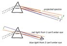
- Take a photo through a glass prism to view a light source. Notice that in the photo taken, the object seen through the prism has two colored edges, one red and one blue (i.e., “dispersion”). Draw a light ray diagram and find out which color has the higher refractive index through the prism. Note that our eyes were seeing a virtual image of the object through the prism.
- Relevant Equations
- ⇒η = sin[(A+D)2] / sinA2
I know that red light has a lower index of refraction than blue light, but that’s not what I’m seeing. The blue light is where the red light should be.
I can’t afford to join CHEGG. Any chance that someone would help me out for free? I’d really appreciate it.
[Link to chegg removed by the Mentors]
I can’t afford to join CHEGG. Any chance that someone would help me out for free? I’d really appreciate it.
[Link to chegg removed by the Mentors]
Last edited by a moderator: