- 7,793
- 4,093
- TL;DR
- petrology
(Edit: since the thread title was changed, this first sentence is too cryptic: the original title referred to a Tool song....)
Besides being a favorite song by a favorite band, the thread title is a straightforward play on words. This summer, as a present to myself for being promoted, I purchased a collection of thin sections that I believe comprise the research materials of Prof. Rob Verschure, who at the time was faculty in the Geological Institute in Amsterdam.
What changed this purchase from eccentric (although, at $2 per sample, also very affordable) to something more elevated is that Prof. Verschure published his findings on many of these samples, primarily in:
https://www.ngu.no/FileArchive/NGUPublikasjoner/NGUnr_380_Bulletin_70_Verschure_35_49.pdf
and
https://research.vu.nl/en/publicati...-parameter-for-metasomatism-and-its-applicati
i.e. I have a "Rosetta stone" for explosion breccias, carbonatites, and damtjernites. Many of the collected samples have been fully characterized, for example this thin section:

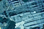
Sample Hor 1 has been classified as a calcite-bearing clinopyroxene-hornblende lamprophyre that has been dated to 313 Ma. This sample contains abundant augite and brown hornblende.
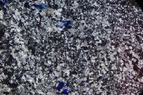
This sample (Fen 23) consists of zoned augite (I think...) and carbonates, dated to 594 Ma:

Many of the samples are carbonatites, but there are also many examples of schists and gneisses; here's a schist with pronounced folding:

I'm hoping to learn quite a bit as I get more familiar with the variety of samples...
Besides being a favorite song by a favorite band, the thread title is a straightforward play on words. This summer, as a present to myself for being promoted, I purchased a collection of thin sections that I believe comprise the research materials of Prof. Rob Verschure, who at the time was faculty in the Geological Institute in Amsterdam.
What changed this purchase from eccentric (although, at $2 per sample, also very affordable) to something more elevated is that Prof. Verschure published his findings on many of these samples, primarily in:
https://www.ngu.no/FileArchive/NGUPublikasjoner/NGUnr_380_Bulletin_70_Verschure_35_49.pdf
and
https://research.vu.nl/en/publicati...-parameter-for-metasomatism-and-its-applicati
i.e. I have a "Rosetta stone" for explosion breccias, carbonatites, and damtjernites. Many of the collected samples have been fully characterized, for example this thin section:
Sample Hor 1 has been classified as a calcite-bearing clinopyroxene-hornblende lamprophyre that has been dated to 313 Ma. This sample contains abundant augite and brown hornblende.
This sample (Fen 23) consists of zoned augite (I think...) and carbonates, dated to 594 Ma:
Many of the samples are carbonatites, but there are also many examples of schists and gneisses; here's a schist with pronounced folding:
I'm hoping to learn quite a bit as I get more familiar with the variety of samples...
Attachments
Last edited: